Inflammation-suppressing cornea-in-a-syringe with anti-viral GF19 peptide promotes regeneration in HSV-1 infected rabbit corneas
Published in Bioengineering & Biotechnology and General & Internal Medicine
Cross-border and trans-Atlantic logistics, and COVID-19. Montreal, Quebec, had the unwanted distinction of being the COVID-19 hardest-hit Canadian city and was completely shut down for months with a very slow re-opening. Our project started with this centre developing biomaterials, nanoparticles and anti-viral peptides to ship to our European partners... A slimmed-down project was finally completed and published as two papers in Scientific Reports (Thathapudi et al. 2024; https://rdcu.be/dB2Hp) and the present paper in npj Regenerative Medicine after several COVID-19-related hiccups!
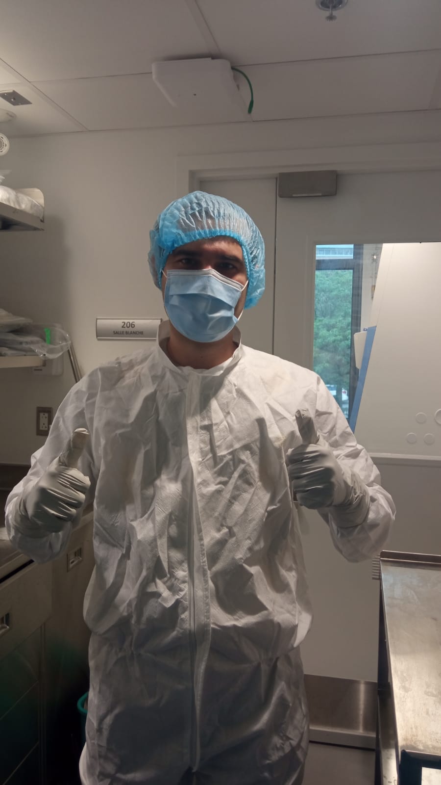
Developing the injectable biomaterials and anti-viral peptides. Our Montreal-based research centre at the Maisonneuve-Rosemont Hospital is fully equipped with a Class 10,000 cleanroom containing a Class 100 hood for biomaterials work. Post-doctoral fellows Elle and Bijay spent many hours developing and optimizing the formulation and figuring out how they would fill the syringes for delivering our "cornea-in-a syringe" or CIS with the injectable hydrogel and the silica nanoparticles containing GF19, a derivative of the cationic innate immune defence peptide, LL37, designed by another post-doctoral fellow, Kamal Malhotra (published in Thathapudi et al., 2024). All three post-docs lost a lot of their time due to COVID. Elle and Bijay left almost right after making up the syringes and shipping them to our partners in Vilnius, Lithuania. Kamal had already left and had not participated in this rabbit study. PhD students, Mostafa, Mozhgan, and Neethi continued the characterization work.

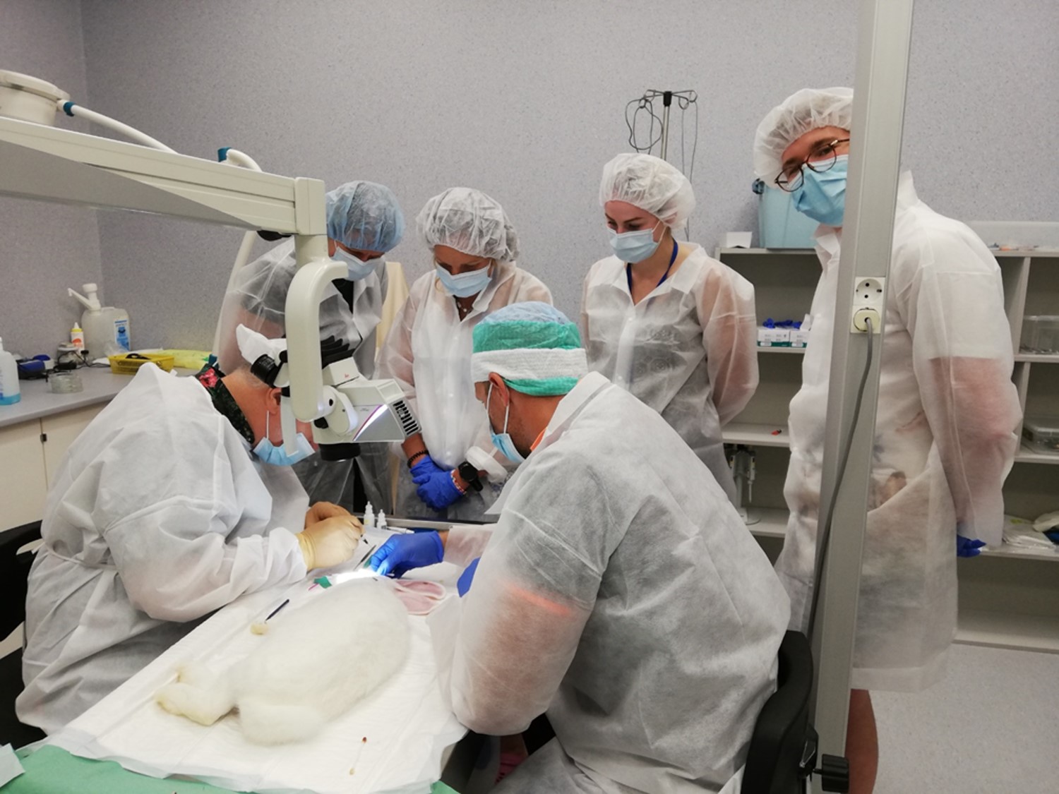
Rabbit cornea surgeries. Ophthalmologists and cornea surgeons, Rimvydas and Almantas, along with Lithuanian project leader Virginija, developed the rabbit HSV-1 infection and surgical perforation model. Egidijus and the rest of the team took turns with the animals' post-operation care, painstakingly noting every minute change in the health of both the treated and control eyes. After ethical permission, the surgeons made a stepped surgical perforation in one cornea of each rabbit, which was filled with cyanoacrylate glue (the standard of care for patients arriving in the emergency room with a perforation in many major centres), CIS alone, or CIS containing GF19 releasing nanoparticles (SiNP-GF19). They made a heroic effort, completing the surgeries of all 24 rabbits over 2 days. Thankfully, CIS was easy to apply and effectively filled the perforations.

Over the recovery and follow-up period, we also noted that compared to cyanoacrylate glue, CIS did not damage surrounding tissues and had much less pronounced scarring. The CIS suppressed inflammation very well. However, we were quite disappointed that the incorporation of SiNP-GF19 in CIS did not demonstrate the inflammation suppression we saw with CIS alone. While analysing tear samples we have observed much less pronounced inflammation and immune response in all CIS formulations compared to cyanoacrylate group. We figured that at least we can say that CIS did better than cyanoacrylate even though SiNP-GF19 was disappointing from the eye exams.
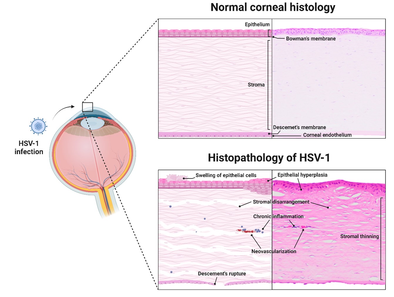
Histopathology and immunohistochemistry. The initial plan was for all the rabbit cornea and lymph node samples to be shipped from Lithuania back to Quebec for histopathology. However, due to COVID-19, trying to get animal samples into Canada was an absolute nightmare. So, it was a change of plans, and in hindsight, extremely fortuitous. Our Spanish partners came to the rescue. Vet pathologists, Jaime and Ines, had to section and examine every cornea and its contralateral control, as well as all the draining lymph nodes. The whole team was very pleasantly surprised when they found that while the CIS with SiNP-GF19 did not appear to do much from the clinical examinations, the histopathology results told a different story. Only the corneas that were given CIS-SiNP-GF19 and additional SiNP-GF19 through an ointment we also made, had morphologies approaching that of normal, healthy rabbit corneas. Then, Yolanda and Miguel who did the immunohistochemistry had an even bigger surprise for the team. They found that HSV-1 viruses had remained within the cells of the cyanoacrylate and CIS-treated and regenerated corneas even at 6 months post-infection, long after the animals stopped shedding virus into their tears. However, the corneas that received CIS-SiNP-GF19 were virus-free. There was no HSV-1 viral staining in these corneas.
Take-home message. Despite the immense challenges posed by COVID-19, our team demonstrated remarkable resilience and dedication, ensuring that we met our deadlines. We truly embodied the spirit of the Beatles' song,' With a Little Help from my Friends', when our Spanish colleagues generously offered their time and support. We look forward to the possibility of future collaborations with current friends and new friends.
And now, some additional information of corneal HSV-1 infections...

Follow the Topic
-
npj Regenerative Medicine

This journal is an open access, online-only, peer-reviewed journal dedicated to publishing high-quality research on ways to help the human body repair, replace and regenerate damaged tissues and organs.
Related Collections
With Collections, you can get published faster and increase your visibility.
Cellular and Genetic Tools in Regenerative Medicine
Publishing Model: Open Access
Deadline: Sep 11, 2026
Heart Regeneration and Beyond-Volume II
Publishing Model: Open Access
Deadline: Jun 01, 2026


Please sign in or register for FREE
If you are a registered user on Research Communities by Springer Nature, please sign in