New imaging pipeline developed to decipher cell-specific metabolic functions
Published in Cancer, Chemistry, and Protocols & Methods
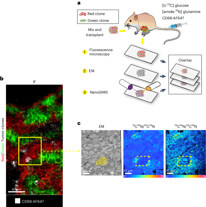
We worked together with collaborators at the Francis Crick Institute and NPL, as part of the CRUK Grand Challenges team Rosetta, and have developed a multimodal imaging pipeline that extends upon the principles of correlative light, electron, and ion microscopy (CLEIM), which combines confocal microscopy reporter or probe-based fluorescence, electron microscopy (EM), stable isotope labelling and Nanoscale secondary ion mass spectrometry (NanoSIMS). Their protocol allows an unprecedented extraction of biological information from specimens, whilst being based on a series of well-established and widely available technologies, thus allowing quick adaptation of the protocol for individual research needs. This integration provides a multifaceted view of the tissue microenvironment, capturing both the internal cellular architecture and the intricate metabolic dynamics occurring within. We tested this pipeline by imaging the incorporation of carbon from glucose into B and T cells in mouse liver tumours.
Read the article here - https://www.nature.com/articles/s41596-024-01118-4
If you don't have a subscription - https://rdcu.be/d7WGO
Follow the Topic
-
Nature Protocols
This journal publishes secondary research articles and covers new techniques and technologies, as well as established methods, used in all fields of the biological, chemical and clinical sciences.


Please sign in or register for FREE
If you are a registered user on Research Communities by Springer Nature, please sign in