Prenatal THC and Long-Term Cognitive/Memory Outcomes
Published in Neuroscience

Understanding the Context:
Cannabis use during conception, pregnancy, and nursing is discouraged by health organizations worldwide. However, in Canada, approximately 22% of pregnant women aged 18-24, report using cannabis [1] . Anxiety, depression, and nausea are the main reasons cited for self-medicating with cannabis during pregnancy [1, 2] . This trend may be attributed to a lack of appropriate counseling from healthcare providers and enduring misconceptions that as a ‘natural product’, cannabis poses little risk to the developing fetal brain. A 2018 maternal cannabis study in Ontario revealed that cannabis use during pregnancy is the third-highest risk factor for low birth weight and long-term health issues. The primary psychoactive component in cannabis, THC, disrupts the endocannabinoid system (ECS) responsible for regulating fetal and adolescent neurodevelopment as well as cognitive and emotional processing. Importantly, over the past two decades, THC concentrations in cannabis have increased from 3% to 22% [3]. THC can cross the placenta, with fetal plasma containing 10-28% of maternal concentrations and fetal tissues exhibiting 2-5 times higher concentrations [4,5,6]. Previous evidence suggests that THC, the primary psychoactive compound in cannabis, can have lasting effects on cognition and lead to lifelong neuropsychiatric outcomes; in humans, the literature strongly suggests that this depends on dose, frequency of use, and duration of use before and during pregnancy, and during lactation [2,7]. Therefore, studying the consequences of THC exposure, in a rodent model, is crucial for understanding the molecular mechanisms underlying these risk factors and identifying intervention strategies to mitigate these potential negative outcomes.
The fetal ECS, which regulates neuronal development and neural circuit refinement, is disturbed by THC. Synaptic signaling events within the ECS rely on plasma membranes composed of esterified fatty acids, which can produce bioactive lipid signaling molecules necessary for ECS function, including the formation of endogenous cannabinoids (endocannabinoids) [8,9]. However, these fatty acids, particularly arachidonic acid (ARA) and docosahexaenoic acid (DHA), are critical for fetal, neonatal, and adolescent neurodevelopment, neuroinflammation, synaptic structure, and function, as well as the regulation of major neurotransmitter systems such as dopamine, glutamate, and GABA [10,11].
Our previous work using rodent models has shown that prenatal THC exposure results in fetal growth restriction (FGR) or low birth weight [4,12,13,14]. Importantly, FGR, regardless of cause, leads to deficient fetal accumulation of polyunsaturated fatty acids (PUFA), particularly the major neural PUFAs, DHA and ARA, which subsequently cause lifelong cardiometabolic and brain health disturbances [4,14,15,16,17]. These PUFAs are essential for ECS function. ARA is converted into anandamide and 2-arachidonoylglycerol, which are vital for ECS neurodevelopmental roles, while DHA is converted into docosahexaenoyl ethanolamine, necessary for neuroinflammatory control and regulating synaptogenesis [18]. Since these PUFAs cannot be synthesized de novo, they are primarily acquired from maternal consumption during the prenatal and neonatal periods. The ECS regulates the acquisition of dietary PUFA precursors and their conversion into free DHA and ARA, which are then transferred to the fetal brain. Insufficient fetal acquisition of these PUFAs during gestation or the neonatal period results in significant structural and functional alterations in the brain. ARA and DHA are integral components of neural cell and synaptic membranes [10,11]. Although prenatal THC exposure disrupts the acquisition of these PUFAs, it does not completely prevent it. However, deficits in PUFA acquisition are associated with disturbances in neurotransmission, neural activity, and cognitive and emotional processing . Further research is needed to determine whether PUFA deficiencies are a cause or consequence of these THC-induced neurobehavioral impairments, as studies have shown that normalizing PUFAs can mitigate neurobehavioral consequences in other prenatal models [19].
Investigating the Effects of Prenatal THC Exposure:
Our study examined the impact of prenatal THC exposure on cognitive function and neurodevelopment. Using a preclinical model, we simulated gestational THC exposure and evaluated its effects on the PFC-hippocampal network, crucial for various cognitive and memory behaviors.
Major Findings:
Our study provides compelling evidence of enduring, sex-selective pathological alterations in the developing male and female brains following chronic THC exposure during gestation. Perhaps most surprisingly, while previous studies have suggested that females are generally more protected against prenatal drug-related insults [14,20,21], both male and female offspring exhibited enduring cognitive deficits in object recognition, social behavior, and working memory. However, these impairments were accompanied by sex-specific sensory filtering deficits, electrophysiological abnormalities, and disturbances in several neurotransmitter systems.
Sex-Specific Effects of Prenatal THC Exposure:
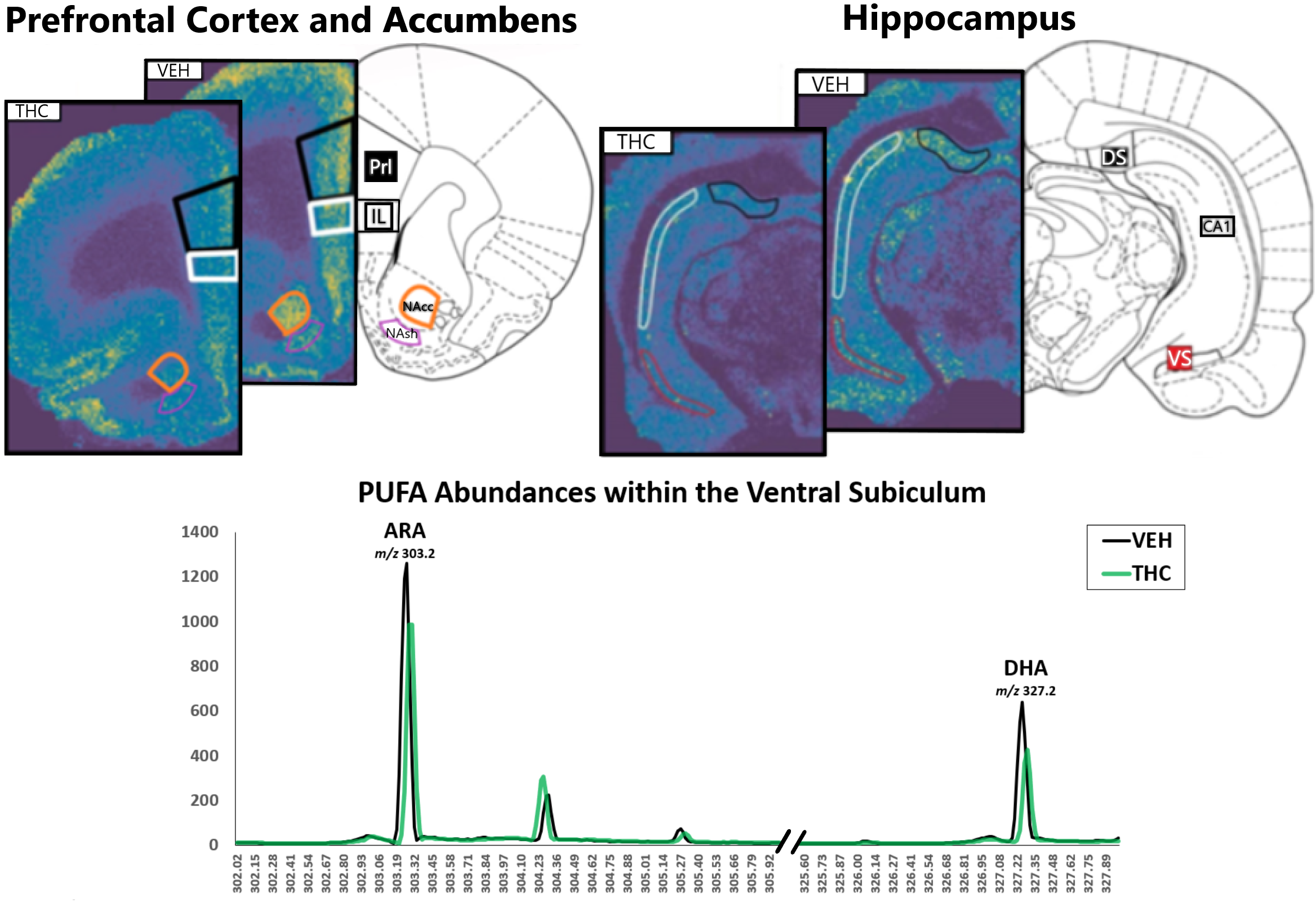
Interestingly, while cognitive deficits were similar between sexes, we discovered that the underlying neuropathological mechanisms differed significantly. For example, females displayed hyperactive neural activity in the ventral hippocampus (vHIPP) and lipidomic deficits in the hippocampal formation. Males, on the other hand, exhibited hypoactive vHIPP activity, oscillatory disruptions in various brain regions, and lipidomic deficits primarily in the PFC (Fig.1; see Figure. 4 in publication). We’ve previously also shown that deficits also persist in the male NAc, while the female NAc is largely protected against both anxiety and these lipidomic abnormalities [14].
Role of Lipidomic Alterations:
Lipidomic changes played a crucial role in the observed pathologies. For example, prenatal THC exposure led to persistent reductions in DHA, ARA, and their metabolites in the PFC and hippocampus. Deficits in these essential fatty acids, crucial for synaptic membrane formation and neural signaling, were linked to disturbances in neurotransmitter systems, including dopamine, glutamate, and GABA. Deficiencies in these neurochemical pathways during key neurodevelopmental periods is likely to lead to severe abnormalities in synaptic structure and function, which we hypothesize may explain many of the observed behavioral and molecular abnormalities. Furthermore, during gestation, deficiencies in PUFAs, the fetal growth restriction (FGR) we’ve previously observed, also can lead to neurocognitive disturbances and negatively impact the frontal lobe and hippocampus. The major departure with these previous FGR studies is that the ECS is disturbed by THC, which potentially exacerbates FGR-related abnormalities, although future studies are required to further examine these pathophysiological processes.

Fig. 1. MADLI-IMS reveals prenatal THC induces cortical lipidome disturbances. VEH vs. THC for DHA in PFC (Prl and Il), NAc (NAcc and NAsh [14]), and hippocampal formation (VS, DS, and CA1).
Sex Hormones and Protective Mechanisms:
Sex-specific effects can be attributed to variations in the ability to convert precursors into DHA and ARA. Females, benefiting from more efficient fatty acid conversion processes and the influence of sex hormones like estrogen, may possess protective mechanisms against THC-induced neurodevelopmental disturbances. Interactions between sex hormones, fatty acid metabolism, and cannabinoid receptors may underlie divergent responses in male and female offspring. Future studies are required to further explore these potential mechanisms.
Implications and Future Directions:
Understanding the lasting effects of prenatal THC exposure on brain development is of crucial importance. Disruptions in lipidomic profiles, neurotransmitter systems and oscillatory patterns contribute to cognitive deficits, altered social behavior, memory impairments, and sensorimotor gating deficits. Targeting lipidomic abnormalities may offer therapeutic interventions to mitigate the adverse effects of prenatal THC exposure. Further research is needed to explore potential strategies for normalization and develop sex-specific approaches to studying THC's effects. In addition, given the phytochemical complexity of cannabis, future studies should consider full-spectrum cannabis exposure effects as well, including cannabidiol and other minor cannabinoids and terpene compounds, all of which might produce differential effects on the developing fetal brain.
Conclusions:
Ours and other studies have conclusively demonstrated that prenatal THC exposure can exert profound and enduring effects on early brain development. Cognitive deficits, disruptions in lipidomic profiles, and alterations in neurotransmitter systems all contribute to the observed pathological phenotypes. Despite prenatal cannabis use being the third highest risk factor for FGR outcomes and associated neuropathological disturbances, there is still ongoing debate regarding its safety in postnatal neurodevelopmental periods. Finally, with increasing trends toward cannabis legalization around the world, there is an impetus to understand the potential sex-specific risks of prenatal THC exposure on fetal development and long-term neurocognitive outcomes. Our translational study is highly innovative for several reasons. First, this study demonstrates that prenatal exposure to THC, which we have established leads to FGR, also results in long-term neurocognitive dysfunction. Second, this is the first study to address how THC alters the cortical lipidome and to identify what effects it may have on long-term neurodevelopmental outcomes. Fetal brain development is arguably the most critical period for determining long-term mental health outcomes and it is therefore imperative to identify potential cannabis-related risks for developing targeted therapeutic interventions and addressing the potential long-term consequences of prenatal cannabis exposure.
Follow the Topic
-
Molecular Psychiatry

This journal publishes work aimed at elucidating biological mechanisms underlying psychiatric disorders and their treatment, with emphasis on studies at the interface of pre-clinical and clinical research.
Your space to connect: The Psychedelics Hub
A new Communities’ space to connect, collaborate, and explore research on Psychotherapy, Clinical Psychology, and Neuroscience!
Continue reading announcement
Please sign in or register for FREE
If you are a registered user on Research Communities by Springer Nature, please sign in