Reprogramming the morphology of colorectal cancer cells in 3D cultures to enhance therapeutic response
Published in Cancer, Protocols & Methods, and Biomedical Research
We have long been fascinated by how cancer cells behave in three-dimensional (3D) environments, especially in response to therapies. Unlike traditional 2D cultures, 3D systems better mimic the architecture and polarity of epithelial tissues – features that are often lost in cancer and linked to poor prognosis and therapy resistance. For example, the morphological epithelial-to-mesenchymal transition (EMT) is a key driver of metastasis and drug resistance in multiple cancers, including colorectal cancer (CRC). Over the years, the Singh lab has studied these phenomena in a piecemeal fashion, using labor-intensive methods that limited throughput and scalability.
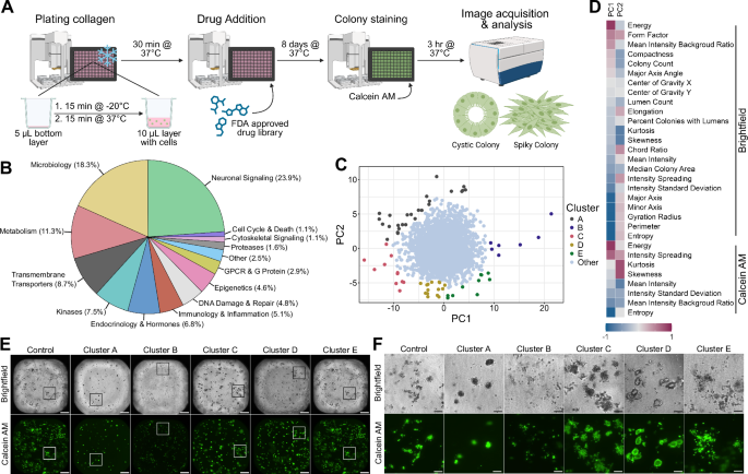
Our new study, Harmych et al. (https://www.nature.com/articles/s42003-025-08699-0), represents a major leap forward. Graduate student Sarah Harmych took on the challenge of miniaturizing these intricate 3D experiments to enable systematic testing of multiple drugs, concentrations, and combinations. In close collaboration with the Vanderbilt High-Throughput Screening Core, we developed a high-throughput drug screening platform employing 3D type I collagen cultures, miniaturizing our approach into a 384-well format while preserving the fidelity and robustness of 3D phenotypes. Using automated imaging and AI-driven analysis, we screened over 1,000 FDA-approved compounds and assessed for their ability to re-epithelialize CRC colonies.
Strikingly, among the top hits were three antibiotics – azithromycin, clindamycin, and linezolid – that consistently induced circular, lumen-containing colonies with restored epithelial features. These morphological changes were accompanied by increased membrane localization of E-cadherin and ZO-1, transcriptomic shifts away from EMT and KRAS signaling, and enhanced sensitivity to the chemotherapeutic irinotecan.
To assess clinical relevance, we conducted a retrospective analysis of CRC patients treated with irinotecan. Those who also received azithromycin had significantly improved 5-year survival rates, suggesting a synergistic interaction between re-epithelialization (EMT reversal) and chemotherapy response.
This study not only identifies promising drug candidates for combination therapy but also introduces a scalable, physiologically relevant platform for morphological drug screening. As the field moves toward human-relevant models and away from animal testing, our 3D collagen-based assay offers a powerful tool for discovering therapies that impact epithelial architecture in overcoming drug resistance.
Follow the Topic
-
Communications Biology
An open access journal from Nature Portfolio publishing high-quality research, reviews and commentary in all areas of the biological sciences, representing significant advances and bringing new biological insight to a specialized area of research.

Related Collections
With Collections, you can get published faster and increase your visibility.
Healthy Aging
Publishing Model: Open Access
Deadline: Dec 31, 2026
DNA repair and human disease
Publishing Model: Hybrid
Deadline: Oct 31, 2026


Please sign in or register for FREE
If you are a registered user on Research Communities by Springer Nature, please sign in