Seeing is believing – how we learned to look at the images
Published in Cancer

I am a 5-th year MD-PhD student at the University of Helsinki, and my research is focused on finding more effective treatments for the common and lethal High-grade serous ovarian cancer (HGSC). HGSC is commonly diagnosed at an advanced stage, and even after extensive surgery and multiple rounds of chemotherapy the disease commonly progresses and leads to devastating outcomes.
Cancer can only develop and progress when the tumor cells develop ways to hide from the body’s immune system. Cancer immunotherapies, which boost the body’s immune defense against cancer, have emerged as promising therapies in multiple tumor types. Unfortunately, immunotherapies have shown poor results in ovarian cancer, and there is an urgent unmet need to find more efficient immunotherapy and combinational therapy strategies for ovarian cancer patients.
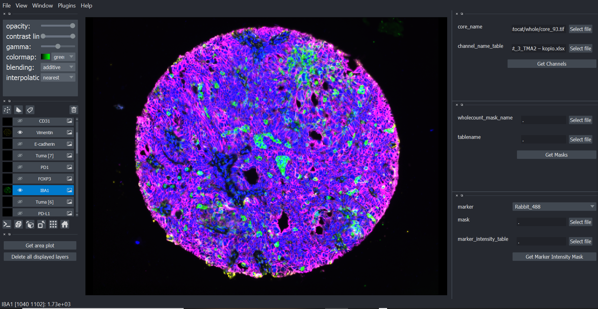
In this work we aimed to investigate whether the genetic alterations shape the tumor microenvironment in HGSC. We compared the tumor-immune microenvironment of BRCA1/2 mutated tumors to those that had no alterations in these genes, or 13 other genes in the homologous recombination pathway. For this,we used a new imaging technology called tissue cyclic immunofluorescence (tCycIF) developed in the Laboratory of Systems Pharmacology led by Professor Peter Sorger at Harvard Medical School. In tCycIF, the fixed tissue sections are stained with antibodies cyclically, so that for each round four to seven fluorescent dyes are used at the same time, the images are scanned with a microscope in small areas called tiles, and then the signal is quenched by bleach and physical light. And then the cycle is repeated, and repeated - up to 20 times. Each staining experiment typically lasts for 2-3 weeks, and after images have been acquired, the real fun - image- and data analysis - begins, and that typically takes 1-2 years. The process starts with stitching the tiles together and aligning the cycles carefully to match each antibody to each single cell in the tissue. The images also need to be preprocessed to remove potential artifacts and uneven signals from the microscope.
Then we do a process called cell segmentation to identify the boundaries of each single cell from the images. In this project we used a machine learning algorithm to identify the nuclei of each cell. For this I first manually labeled thousands of nuclei, membranes, and background area to train the algorithm. Tumor cells with polymorphic nuclei, or cells close to each other and partially overlapping caused trouble - sometimes even the human eye could not distinguish between different nuclei, so how can we make sure that the algorithm can? The solution to this was only to do more and more careful manual labeling and repeat the segmentation. Even though cell segmentation can never be perfect, we finally managed to obtain reliable results! The key to success was to really keep looking at the images, the cells, and the segmentation masks.
The next step was to annotate the cells using the quantified signal from the 21 markers. As we had highly multiplexed data with 21 marker expression and morphological data from the cells, we quickly realized that traditional gating of negative and positive cells would not work. In these types of data, each cell phenotype is characterized by an alternating combination of the different marker expressions, and the cells were impossible to annotate by using only one or two markers by gating or visually from the images. To address this, we used a clustering approach in an iterative way in the brilliant CYTO tool developed by a postdoc Julia Casado in our Lab.
The cell type annotation turned out to be a challenge, but at the same time it gave us new insights into the characteristics of the cells and how we can best identify them. We noticed that relying only on clustering and looking at cluster properties resulted in less-than-ideal results – after looking at the cells in the tissues we could see that many of them had been misclassified. We found that distinguishing the immune cell subpopulations from each other was especially challenging due to the spillover of signal from the closely adjacent cells, which is a fairly common phenomena in microscopy image-based single-cell data. We solved this issue again by projecting the segmentation masks of the annotated cell types on top of the microscopy images which enabled us to visually confirm the existence of the distinct phenotypes. Also, when cells that we could not annotate using clustering were projected into the images, we were able to classify these cells into meaningful cell types on many occasions. I also think that it is crucial to further investigate these cells that remain unclassified using only prior knowledge – there lies a great potential to discover completely new cell phenotypes! In total we were able to classify the immune cells into ten different cell types, and the tumor and stromal cells further into functional subpopulations.
Based on my experience I can say that seeing is believing - only by carefully and repeatedly looking at the images we can be confident in the cell segmentation and cell type annotations. Using this approach, I was able to fully dive into the analysis of the single-cell spatial data and uncover the unique tumor microenvironment in HGSC.
Follow the Topic
-
Nature Communications
An open access, multidisciplinary journal dedicated to publishing high-quality research in all areas of the biological, health, physical, chemical and Earth sciences.

Related Collections
With Collections, you can get published faster and increase your visibility.
Women's Health
Publishing Model: Hybrid
Deadline: Ongoing
Tumor Microenvironment Crosstalk and Therapeutic Implications
Publishing Model: Hybrid
Deadline: Nov 02, 2026
Please sign in or register for FREE
If you are a registered user on Research Communities by Springer Nature, please sign in