Smokeless tobacco (Shammah) extract: effects on hematological parameters, antioxidant defense mechanisms, and organ health in rats
Published in Chemistry, Biomedical Research, and Agricultural & Food Science
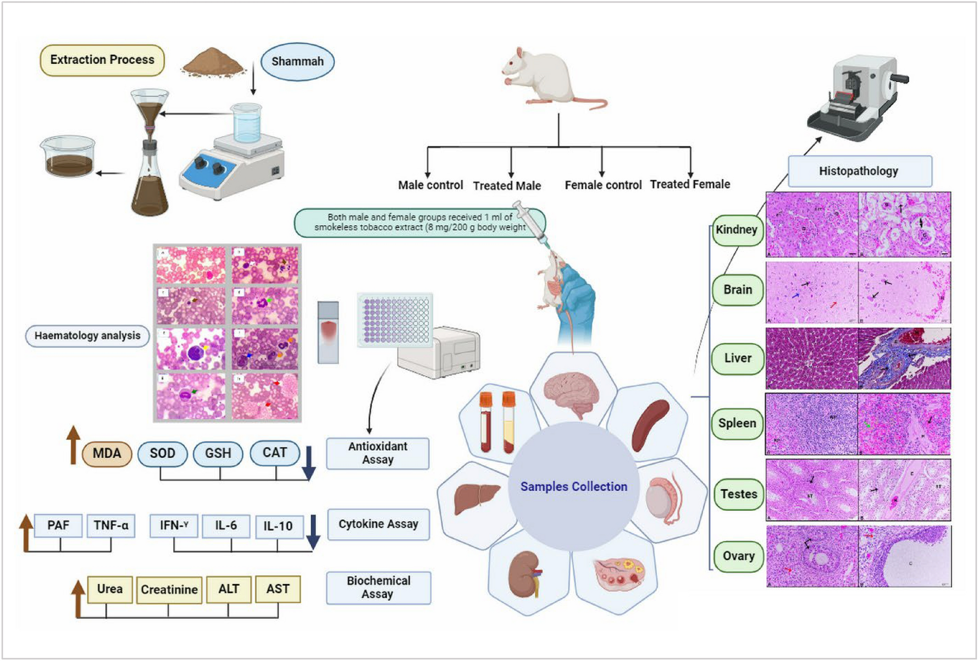

The administration of Shammah extract inducedhematological, biochemical, and histopathological changes in female and male rats. Treated females showed a decrease in total leukocyte count (TLC) to 9900, while treated males increased to 14,525. Lymphocyte percentage decreased by 9.5% in females and 6.02% in males, with neutrophil counts rising by 24.6% and 20.5%, respectively. Eosinophil levels surged by 240% in females and 50.3% in males. Hemoglobin levels decreased by 12.4–13.1% in females, while males showed a non-significant increase to 15.68. Malondialdehyde (MDA) levels increased to 1.57 in females (57% increase) and 1.93 in males (70.8% increase). Antioxidant enzymes decreased, with superoxide dismutase (SOD) at 3.53 (116.2% decrease) in females and 3.90 (45.8% decrease) in males. Kidney function assessments revealed elevated urea levels of 36.35 (84.8% increase) in females and 43.17 (131.2% increase) in males, alongside creatinine levels of 1.28 (75.3% increase) in females and 1.56 (90.2% increase) in males. Histopathological examinations showed untreated livers with a typical structure, while treated livers exhibited infiltrative cell aggregations, venous congestion, hemorrhage, and edema. Treated kidneys showed severe glomerular atrophy and degeneration. Spleens from treated groups had blending of white and red pulp, while brains displayed hemorrhage and distorted neurons in males, and ghost neurons in females. Treated testes exhibited dilated blood vessels, edema, and reduced spermatogenesis, while treated ovaries showed cyst formation and vacuolar degeneration. Our findings indicate significant oxidative stress and organ damage associated with Shammah extract exposure.
 https://link.springer.com/article/10.1007/s10735-025-10403-9
Mohamed Fawzy Ramadan is a Professor of Biochemistry at the Department of Clinical Nutrition, Faculty of Applied Medical Science, Umm Al-Qura University, Makkah, Saudi Arabia.
Prof. Ramadan obtained his Ph.D. (Dr. rer. nat.) in Food Chemistry from the Berlin University of Technology (Germany, 2004). Prof. Ramadan continued his postdoctoral research at ranked universities, including the University of Helsinki (Finland), the Max Rubner Institute (Germany), the Berlin University of Technology (Germany), and the University of Maryland (USA). In 2012, he was appointed as a Visiting Professor (100% teaching) in the School of Biomedicine, Far Eastern Federal University in Vladivostok, Russian Federation.
Prof. Ramadan has published more than 450 research papers and reviews in international peer-reviewed journals. He also edited and published several books and book chapters (Scopus h-index is 55 and more than 11000 citations). He was an invited speaker at several international conferences. Since 2003, Prof. Ramadan has been a reviewer and editor for several highly cited international journals, including the Journal of Medicinal Food, eFood, the Journal of Umm Al-Qura University for Applied Sciences, and the Journal of Advanced Research. He is the editor-in-chief of Journal of Umm Al-Qura University for Medical Science.
Prof. Ramadan received several prizes, including Abdul Hamid Shoman Prize for Arab Researcher in Agricultural Sciences (2006), the Egyptian State Prize for Encouragement in Agricultural Sciences (2009), European Young Lipid Scientist Award (2009), AU-TWAS Young Scientist National Awards (Egypt) in Basic Sciences, Technology and Innovation (2012), TWAS-ARO Young Arab Scientist (YAS) Prize in Scientific and Technological Achievement (2013), and Atta-ur-Rahman Prize in Chemistry (2014).
Follow the Topic

Related Collections
With Collections, you can get published faster and increase your visibility.
SDG 3 – Reducing Global Maternal Mortality
Publishing Model: Hybrid
Deadline: Ongoing