Sugar-coated Cells for Improved Regenerative Therapies
Published in Bioengineering & Biotechnology and Materials
Upon transplantation of regenerative cell therapies, adhesion is the first and most critical step towards the functional integration of therapeutic cells. Transplanted cells must arrive at the site and successfully anchor themselves long enough to engraft and function. However, many therapeutic cells which show promise for repair and regeneration frequently fail to adhere robustly after transplantation. Genetic modification has classically been a cornerstone strategy for engineering cell surfaces with non-native moieties, the principal example being, chimeric antigen receptors of CAR-T cells. This approach allows for the expression of surface proteins, but requires complex manufacturing, poses regulatory challenges, and carries inherent mutative risks. Therefore, our team became interested in tackling the problem of poor engraftment from a chemical-engineering perspective.
Inspired by the role that polysaccharides play within wet adhesion in vivo, we explored the use of polysaccharide biomaterials as surface functionalities, specifically hyaluronic acid (HA) and alginate (Alg). We hypothesised that these ‘sweet and sticky’ sugar polymers might aid adhesion by facilitating multimodal mechanisms of hydrogen bonding, Van der Waals and Ca2+ coordination between the surfaces of transplanted cells and endothelial cells.
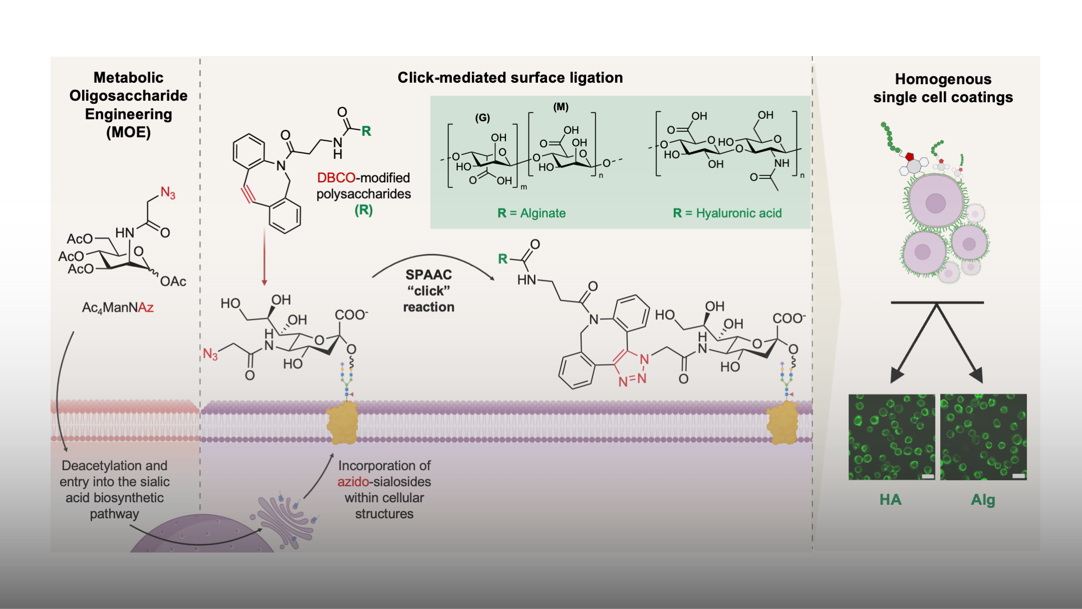
Building on the seminal work of Prof. Bertozzi and colleagues, we adopted metabolic oligosaccharide engineering (MOE) as a method to attach these biopolymers to regenerative cells. This involves the installation of azide anchors on terminal surface sialosides of hepatic progenitor cells (HPCs), through which we were able to chemically ‘click’ adhesive polysaccharides at the cell surface (Fig. 1). This non-genetic approach expands the plethora of potential surface modifications for cell therapy to non-canonical gene products such as synthetic and natural macromolecules, as used in our study.
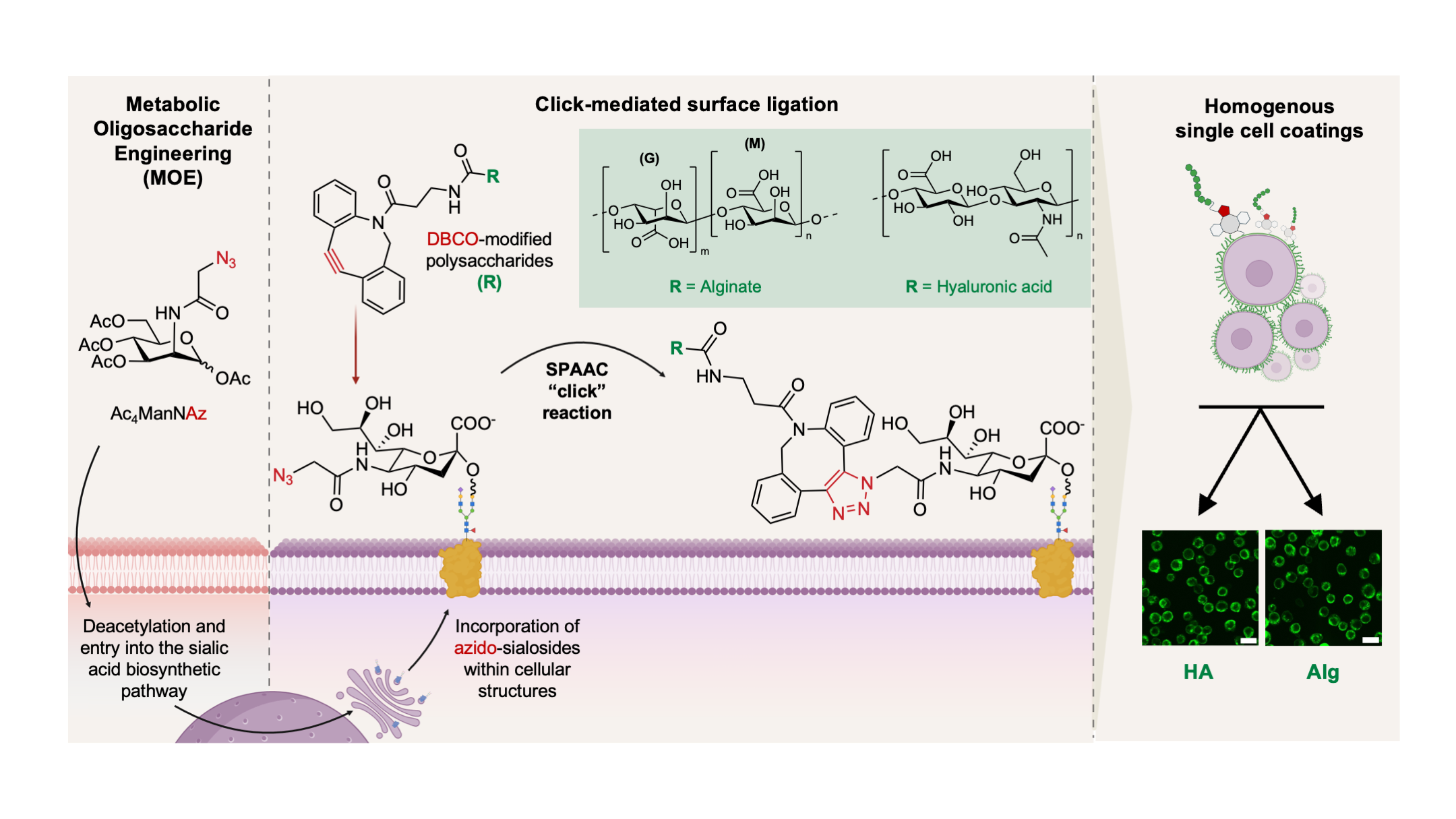
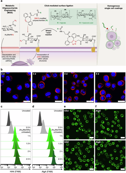
Figure 1. Schematic of metabolic glycan labelling of mammalian cells resulting in the incorporation of N-azidoacetyl-D-neuraminic acid (NeuAz) on surface sialosides, to which, we attach DBCO-modified hyaluronic acid (HA) and alginate (Alg) to achieve cell surface functionalized cells. Confocal fluorescence images of hepatic progenitor cells functionalized with hyaluronic acid (HA) and alginate (Alg) shown in bottom right. Scale bar = 50 mm.
One of the main reasons for using MOE over other surface modification methods such as ionic layer by layer deposition or hydrophobic membrane insertion is that MOE can achieve robust covalent linkage in a highly specific and biocompatible manner. We considered covalent attachment an important factor in our applications as it could resist potential shear stresses upon delivery. Additionally, we adopted this strategy in recognition of the transient nature of the exogenous functionalities at the cell surface, which are progressively lost due to active membrane remodelling and glycan turnover. Indeed, we demonstrate that both Alg and HA coatings were fully lost within a period of ~ 3 days (Fig. 2). This transient display is advantageous within cell therapy applications, wherein the attached functionalities may be subsequently cleared following initial adhesion, allowing the cells to revert back to their native functional phenotypes.
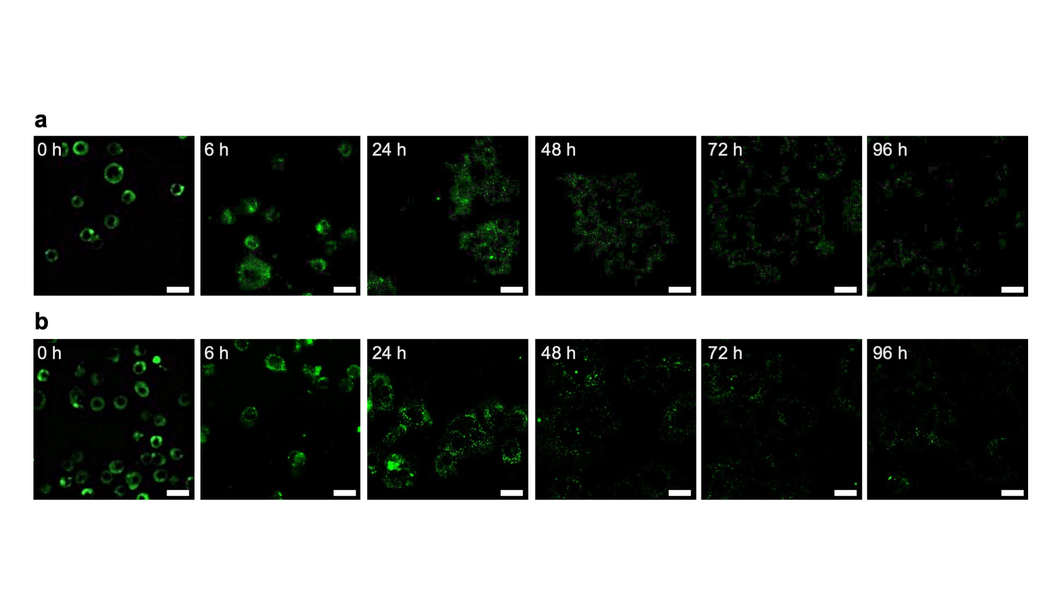
Figure 2. Confocal fluorescence microscopy images of HPCs in standard cell culture media after coating with HA (a) and Alg (b) captured over a period of 96 h. Scale bar = 20 µm.
After establishing a method for robust, single-cell surface coatings, we then probed how these modifications may influence cell adhesion to physiologically relevant surfaces, beginning with model extracellular matrix (ECM) substrates, progressing to 2D endothelial and 3D in vitro liver models. We initially anticipated subtle differences between the Alg and HA, but instead found striking contrasts between the two functional biomaterials. HA-coated cells showed increased levels of adhesion and also showed pronounced morphological features of adhesion (i.e. active membrane remodelling indicated by spreading and protrusions) on both ECM surfaces and 2D HUVEC monolayers, likely as a result of the native functional interactions of HA. Indeed, this supports previous studies in which the upregulation of endogenous HA synthases led to similar phenotypes; seeing such cellular behaviour induced non-genetically was both validating and exciting in our efforts. In contrast, Alg-coated cells responded in the opposite way, remaining rounded and exhibiting reduced spreading, despite being exposed to the same environments. We suspected that this was due to the formation of a relatively stiff alginate layer at the cell surface, caused by Ca2+-mediated crosslinking at the cell surface, effectively encasing the cells in a thin hydrogel shell and preventing cellular interaction with the ECM and endothelial surfaces.
Integrins are the primary surface proteins involved in cellular adhesion, so we wanted to understand how the attachment of these biopolymers affect integrin expression after coating. Through RT-qPCR, we observed something unexpected yet promising for our application. Coated cells showed a broad upregulation in integrin subunits, namely α1, α2, α5, α6, αv and β3, β4, β5. At first glance, we thought this may have been due to technical or biological variability, however, repeated experiments confirmed a consistent trend. Our prior hypothesis of alginate crosslinking at the cell surface led us to explore the literature surrounding mechanobiology, suspecting that the coatings were actively modulating how cells sense their environment. Increase in gene expression of such subunits aligns with known stiffness-induced mechanotransduction pathways, suggesting that the presence of covalently tethered biopolymers may sensitise cells to mechanical forces which native, uncoated cells would not usually perceive. While detailed molecular mechanisms remain to be explored, our results suggest a dual role for our strategy, in which the coatings play an active part in adhesion whilst also priming cells for integrin-mediated engraftment post-transplantation.
After the promising adhesive behaviour of HA-coated cells on 2D models, we then explored whether these effects would translate to more physiologically relevant conditions. We aimed to simulate the conditions that transplanted cells face during engraftment in vivo by incorporating dynamic flow and liver-specific microtissues into our model. To this end, we used a gravity-based microfluidic flow chip developed by our collaborators at InSphero AG in which 3D InSight™ human liver microtissues were used as a model for the liver parenchyma (fig. 3a). After 9 h of flow, coated cells showed a nearly 4-fold increase in adhesion of HA-coated cells compared to uncoated controls (fig. 3b).
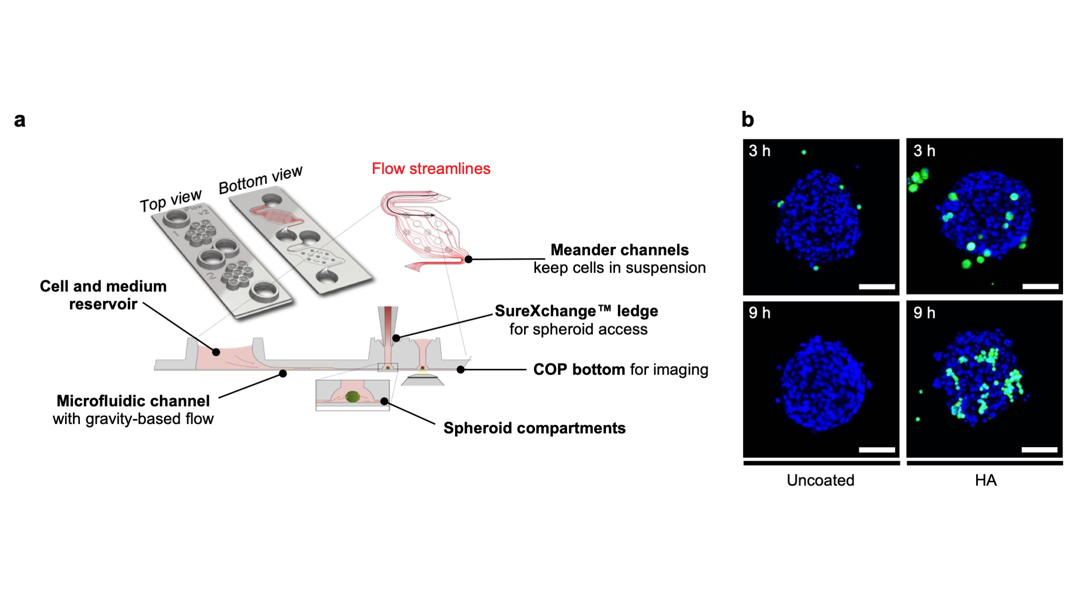
Figure 3. Schematic representation of InSphero AkuraTM ImmuneFlow chip used for the study of coated HPCs engraftment to hLMTs under gravity-induced flow. b. Representative confocal fluorescence images of uncoated and HA-functionalized HPCs (green) engrafted to hLMTs after 3 h and 9 h of incubation under continuous flow. Scale bar = 100 µm
With these results, a critical question that followed was whether improving adhesion might inadvertently compromise cell function. Encouragingly, we demonstrated that the coated HPCs maintained their differentiation capacity, evidenced by phenotypic function after wnt-induced differentiation. Overall, the findings from this study demonstrate this approach as a viable method for improving therapeutic cell attachment, opening up exciting possibilities for translation to other cell types and using new functional materials to develop next generation cell therapies.
To read our full paper in more detail, please follow this link: 'Sweet and sticky: increased cell adhesion through click-mediated functionalization of regenerative liver progenitor cells'
Follow the Topic
-
Communications Biology
An open access journal from Nature Portfolio publishing high-quality research, reviews and commentary in all areas of the biological sciences, representing significant advances and bringing new biological insight to a specialized area of research.

Related Collections
With Collections, you can get published faster and increase your visibility.
Healthy Aging
Publishing Model: Open Access
Deadline: Dec 31, 2026
DNA repair and human disease
Publishing Model: Hybrid
Deadline: Oct 31, 2026


Please sign in or register for FREE
If you are a registered user on Research Communities by Springer Nature, please sign in