Taming the optical resolution, depth of focus, and chromatic aberration of a super-oscillatory lens with genetic algorithm for multicolor super-resolution microscopy
Published in Protocols & Methods
Super-oscillatory lens (SOL) has emerged as a promising diffractive optical element to overcome the resolution limit of objective lenses dictated by the well-known Rayleigh criterion [1, 2]. Recently, SOL-integrated optical microscopes [3, 4] have achieved super-resolution imaging well beyond the diffraction limit, providing an alternative far-field, label-free, non-invasive super-resolution imaging method. However, they still suffer from several intractable issues in practical application scenarios, including 1) inevitable side-lobes, 2) limited depth of focus (DOF), 3) significant chromatic aberration, and 4) short working distance (WD). These issues have imposed significant challenges in commercial realization of SOL-empowered multicolor super-resolution optical microscopy. To date, only one or two of these issues can be tamed successfully with the SOLs reported in the literature [7-15].
How to address the above mentioned challenges has encompassed not only all my Ph.D. journey that concluded in 2021 at the Northwestern Polytechnical University but also the subsequent post-doctoral training in the same institution [5, 6]. During this unforgettable period of 6 years, I have accumulated invaluable experience in applying genetic algorithms for optical designs and the mass fabrication of binary-phase masks with parallel optical lithography, which all have turned out to benefit the ultimate realization of powerful SOLs.
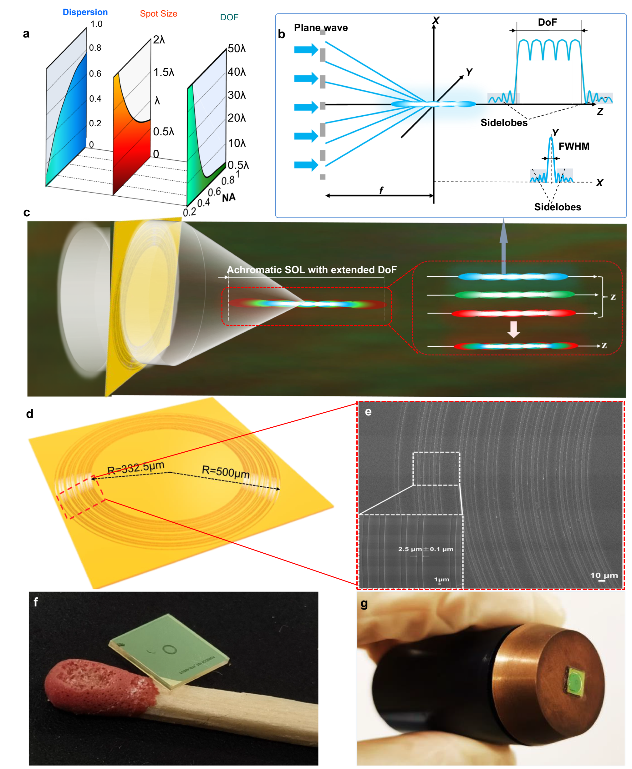
Based on the above experience, my teammate and I have developed a universal method to optimize the key performance parameters of SOLs. Our efforts have finally been materialized in the form of an apochromatic binary-phase SOL, which is designed and optimized by a customized multi-objective genetic algorithm (GA) approach (see Fig. 1). The resultant lens design exhibits an extraordinary imaging capability: it can produce an apochromatic, deep-subwavelength-thin, needle-like focal region with an extended DOF and an augmented WD within the far-field realm, outperforming the best planar lenses with high numerical apertures (NAs) reported in the literature [1, 4].

Fig. 1 a NA-dependent tradeoff among the chromatic dispersion, focusing spot size and DoF of a diffractive lens such as a planar SOL. b Conceptual formation of a needle-like optical pattern by connecting adjacent multifoci under monochromatic illumination, with the minimized side-lobe intensity and main-lobe FWHM. c Schematic illustration of sub-diffraction-limit focusing by an apochromatic SOL with an extended DoF over the whole visible spectrum. The inset sketches the formation of an apochromatic sub-diffraction-limit optical needle by superposing the focus contours at the blue, green and red light wavelengths.
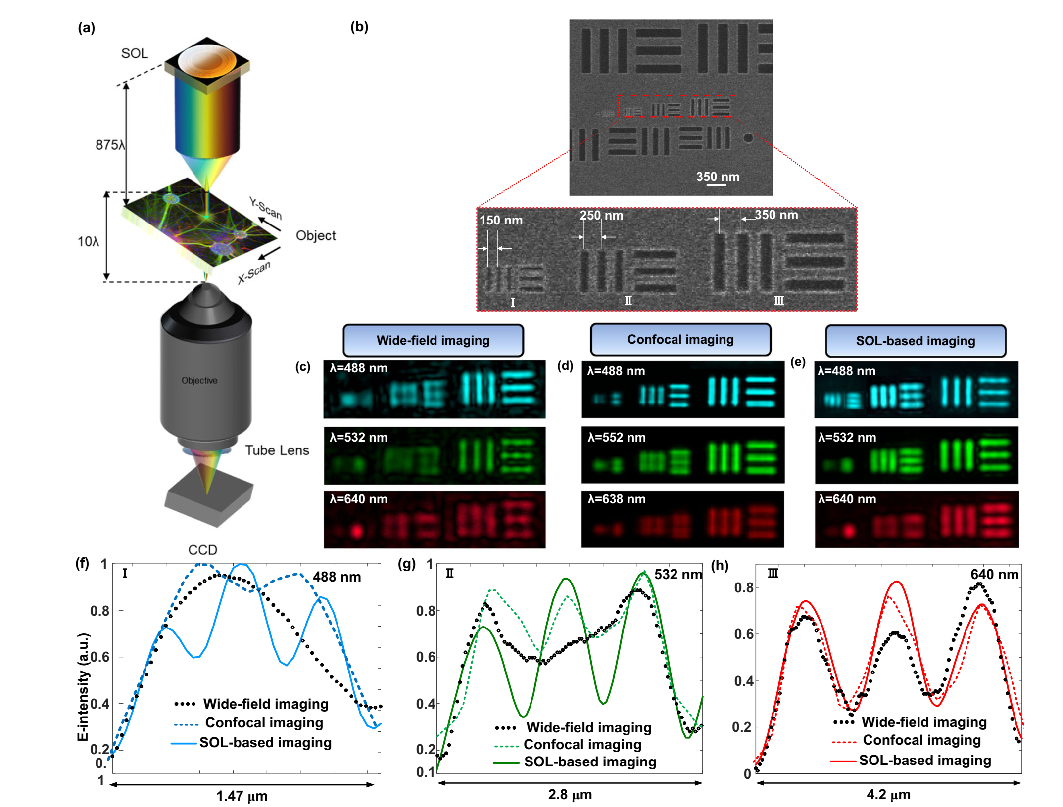
The thinned optical needle and the apochromatic feature of our GA-optimized SOL bring about an amazing opportunity for realizing multicolor super-resolution imaging within the far-field domain, which is achieved by overlapping the subwavelength-thin optical needles of blue, green, and red beams in the same focal region. In experiment, we have integrated this powerful SOL device with a standard fluorescence microscope. Thanks to the extended DOF, we are able to demonstrate label-free super-resolution imaging of 3D objects without axial scanning (see Fig. 2). More remarkably, we have for the first time demonstrated multicolor super-resolution imaging of 3D neuron cells with multiple fluorescent labelling at one go (see Fig. 3). Notably, the far-field resolution limit of our SOL-integrated optical microscope is 0.3λ, far below the super-resolution criterion of 0.5λ/NA.

Fig. 2 Axial-scanning-free imaging of a 3D object. a Sketch of a 3D metallic fishnet wedge composed of an array of circular holes with a period of 2 μm. The depth of the wedge varies from 0.05 μm at the left edge to 1.3 um at the right edge, and the diameter of the holes is about 500 nm. b Top-view (x-y plane) SEM micrograph of a fabricated fishnet wedge. The two insets at the bottom are the zoom-in images of a single hole (left panel) and the top-right corner of wedge. c–e Images of the wedge in b collected by the wide-field microscope c, LSCM d, and our DOF-extended SOL-based microscope all working in the transmission mode (T-mode).

Fig. 3 3D dual-color cellular imaging. a Bright-field and b, c Wide-field fluorescence microscopy images of a thick neuron tissue. e, f Zoom-in views of the enclosed areas in b, c. h, i SOL-based microscopy images of the enclosed area in b, c. d, g Merged dual-color fluorescence images from wide-field imaging and SOL-based microscope. The red arrows in e, f, h, i) denote the fine structures of neurons.
Undoubtedly, scientific research and technological innovation are never smooth sailing, especially when facing the challenges posed by the COVID-19 pandemic. Great thanks to our collaborators, Prof. Lei (one of the corresponding authors) and Dr. Fan (the fourth author) from CityU, who both played an instrumental role in unearthing the significance of our method, designing experiments and also refining the manuscript writing. I hold profound gratitude to Prof. Li (the 8th author), Prof. Yuan and Dr. Liu (the 9th author) for their invaluable assistance in fabricating the complex 3D fishnet wedge (as shown in Fig. 2), which was one of the biggest challenges to demonstrate the superiority of our SOL-based microscopy in label-free super-resolution imaging of 3D objects. I believe that all of our findings presented in this work are the best reward to our persistent endeavors during the past challenging period. I cherish it.
With a deep sense of conviction, we assert that our innovative methodology, featuring flexible customization of SOL optical contours, has effectively surmounted the inherent contradiction between high NA, chromatic aberration, large DOF, and long WD for traditional objective lenses. Besides, the side-lobe intensity of the optical needle can be purposely tuned to accommodate diverse requirements of biological samples for super-resolution fluorescence imaging yet without damaging the biological activity. Such harmonious fusion of technical precision and biological sensitivity underscores the sophistication of our approach. We believe that the demonstrated high-NA apochromatic SOL-based imaging modality with extended DOF and ultra-long WD successfully extends the application scope of optical microscopes to regimes that cannot otherwise be achieved by commercially available, complicated and costly microscope systems.
In summary, our work marks a transformative leap in optical imaging technology, redefining the boundaries of what can be achieved and setting the stage for a new generation of versatile, efficient, and accessible imaging tools with far-reaching applications.
References:
[1]. Chen, , Wen, Z., and Qiu, C. Superoscillation: from physics to optical applications. Light Sci. Appl. 8, 1 (2019).
[2]. Zheludev, N. I. and Yuan, G. Optical superoscillation technologies beyond the diffraction limit. Nat. Rev. Phys. 4, 16 (2022).
[3]. Qin F., Huang, K., Wu, J. A supercritical lens optical label-free microscopy: sub-diffraction resolution and ultra-long working distance. Adv. Mater. 29(8), 1602721 (2017).
[4]. Yuan, G., Rogers, E. T. F., and Zheludev N. I. Achromatic super-oscillatory lenses with sub-wavelength focusing. Light Sci. Appl. 6(9), e17036 (2017).
[5]. Li, W., Yu, Y., Yuan, W. Flexible focusing pattern realization of centimeter-scale planar super-oscillatory lenses in parallel fabrication. Nanoscale 11, 311-320 (2019).
[6]. Li, W., He, P., Yuan, W., Yu, Y. Efficiency-enhanced and sidelobe-suppressed super-oscillatory lenses for sub-diffraction-limit fluorescence imaging with ultralong working distance. Nanoscale 12, 7063-7071 (2020).
[7]. Li, M., Li, W., Li, H., Zhu, Y., Yu, Y. Controllable design of super-oscillatory lenses with multiple sub-diffraction-limit foci. Sci Rep. 7, 1335 (2017). 487
[8]. Huang, K., Ye, H., Teng, J., Yeo, S. P., Luk'yanchuk, B., Qiu, C. Optimization-free superoscillatory lens using phase and amplitude masks. Laser Photon. Rev. 8, 152-157 (2014).
[9]. Rogers, K. S, Bourdakos, K. N, Yuan, G., Mahajan, S., Rogers, E. T. F. Optimising superoscillatory spots for far-field super-resolution imaging. Opt. Express. 26, 8095-8112 (2018).
[10]. Tang, D., et al. Ultrabroadband superoscillatory lens composed by plasmonic metasurfaces for subdiffraction light focusing. Laser Photon. Rev. 9, 713-719 (2015).
[11]. Wu, Z., et al. Broadband dielectric metalens for polarization manipulating and superoscillation focusing of visible light. ACS Photonics 7, 180-189 (2020).
[12]. Yu, Y., Li, W., Li, H., Li, M., Yuan, W. An investigation of influencing factors on practical sub-diffraction-limit focusing of planar super-oscillation lenses. Nanomaterials 8, 185 (2018).
[13]. Chen, G., et al. Super-oscillatory focusing of circularly polarized light by ultra-long focal length planar lens based on binary amplitude-phase modulation. Sci Rep. 6, 29068 (2016).
[14]. Ye, H., Qiu, C., Huang, K., Teng, J., Yeo, S. P. Creation of a longitudinally polarized subwavelength hotspot with an ultra-thin planar lens: Vectorial rayleigh-sommerfeld method. Laser Phys. Lett. 10, 065004 (2013).
[15]. Zhang, S., et al. Synthesis of sub-diffraction quasi-non-diffracting beams by angular spectrum compression. Opt. Express. 25, 27104-27118 (2017).
Follow the Topic
-
Nature Communications

An open access, multidisciplinary journal dedicated to publishing high-quality research in all areas of the biological, health, physical, chemical and Earth sciences.
Related Collections
With Collections, you can get published faster and increase your visibility.
Women's Health
Publishing Model: Hybrid
Deadline: Ongoing
Advances in neurodegenerative diseases
Publishing Model: Hybrid
Deadline: Mar 24, 2026


Please sign in or register for FREE
If you are a registered user on Research Communities by Springer Nature, please sign in