Three-dimensional inner fracture insights into the complex fracture behavior of superalloys
Published in Bioengineering & Biotechnology, Materials, and Computational Sciences
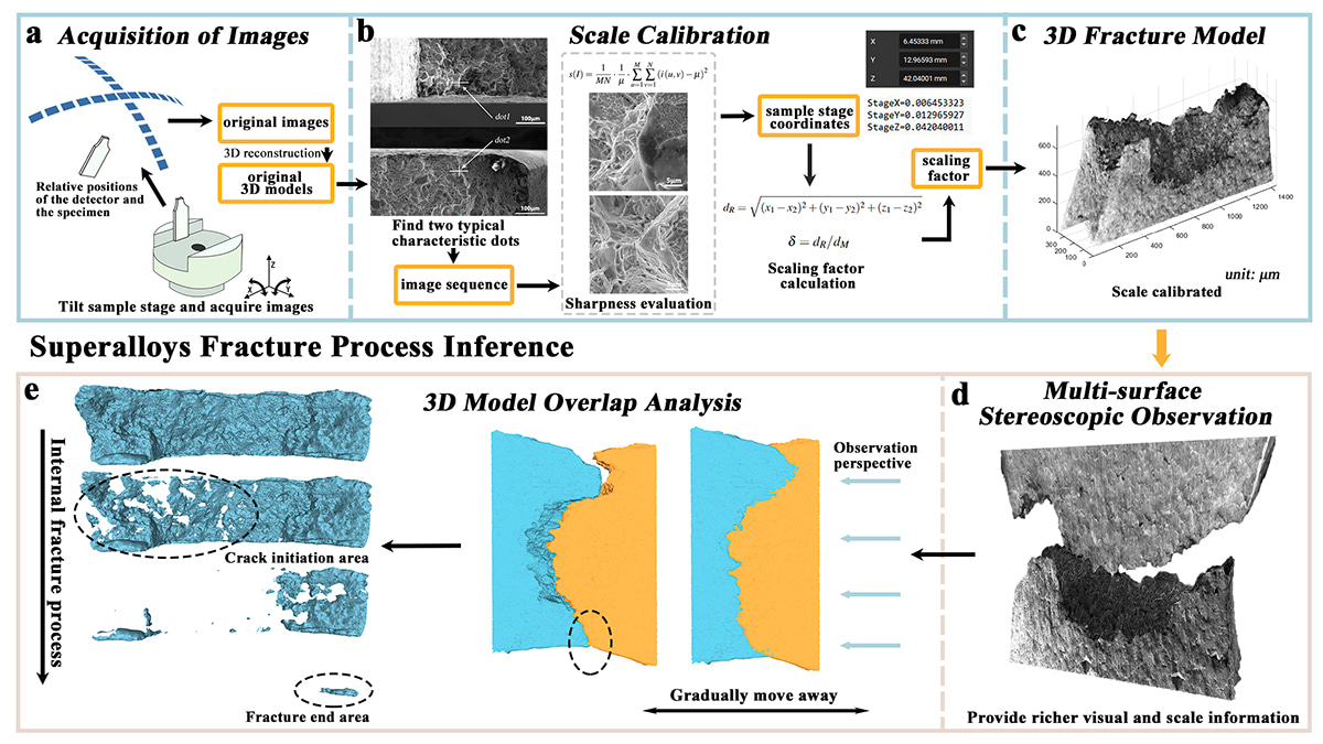
As engineers and material scientists, we are often captivated by the invisible: the microscopic world where materials' strength is defined and failures initiate. Our recent publication in Communications Engineering, "Superalloys fracture process inference based on overlap analysis of 3D models," delves into this realm, aiming to demystify the inner fracture processes of superalloys, materials critical to aerospace and energy industries due to their remarkable properties at high temperatures and pressures.
The Inspiration Behind Our Study
The genesis of this research was sparked by a fundamental challenge in material science: understanding how and why superalloys fracture. Traditional analysis methods provide a snapshot of fracture surfaces but offer limited insight into the dynamic fracture process. In-situ thermo-mechanical deformation experiments can only observe information from a single surface, and it is impossible to know the cracking and crack propagation on the inside or the back of the sample, which brings significant limitations. If one wants to see the evolution of information inside, it is necessary to combine in-situ CT technology, at which point the morphological characteristics brought by SEM will be lost. Therefore,we sought to develop a methodology that could peer into the material's internal deformation, offering a more comprehensive view of fracture initiation and propagation.
The Journey of Discovery
Our journey began with the selection of IN718 superalloy, a widely used material in aerospace applications. We designed in situ tensile experiments to observe the material's behavior under stress. The experiments were performed at room temperature and at an elevated temperature of 650°C to capture the material's response under varying thermal conditions.
To reconstruct the 3D fracture morphology, we employed an optimized image acquisition method, capturing images from multiple angles using a scanning electron microscope (SEM), as Figure 1 shown. This approach was pivotal as it allowed us to create detailed 3D models that could be virtually dissected to analyze the fracture process.
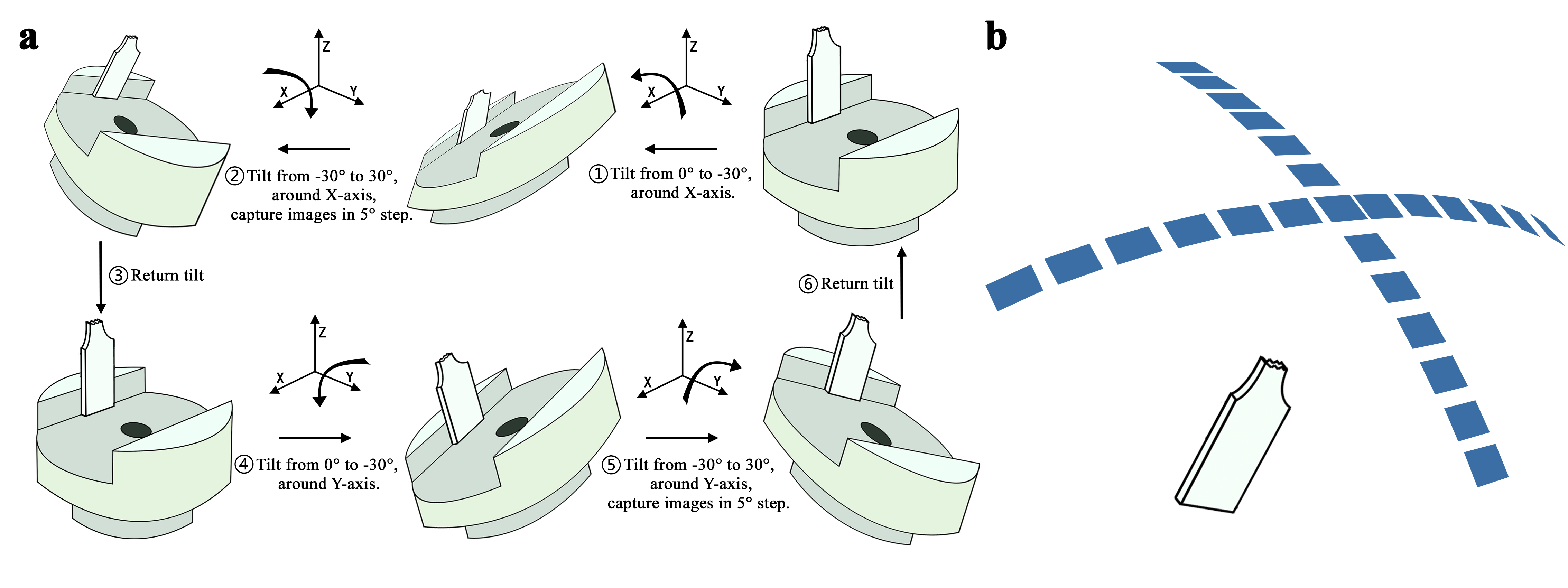
Figure 1. Optimized scheme for original image acquisition. a Detailed tilting steps of the sample stage. b Relative positions relationship between detector and specimen.
Innovative Approaches and Methodologies
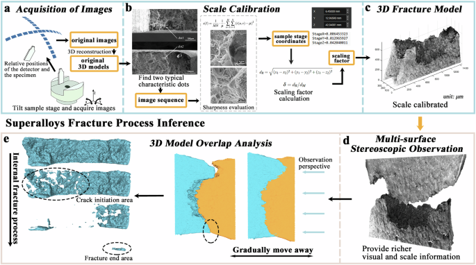
We develop a superalloys fracture process inference (SFPI) framework consisting of multiple steps (Figure 2). The crux of our innovation lies in the 3D model overlap analysis, a method that infers the fracture process by virtually recombining the fractured surfaces. This technique revealed that material deformation and fracture likely result from a combination of surface and internal cracking, challenging the traditional view of fracture as an isolated event.
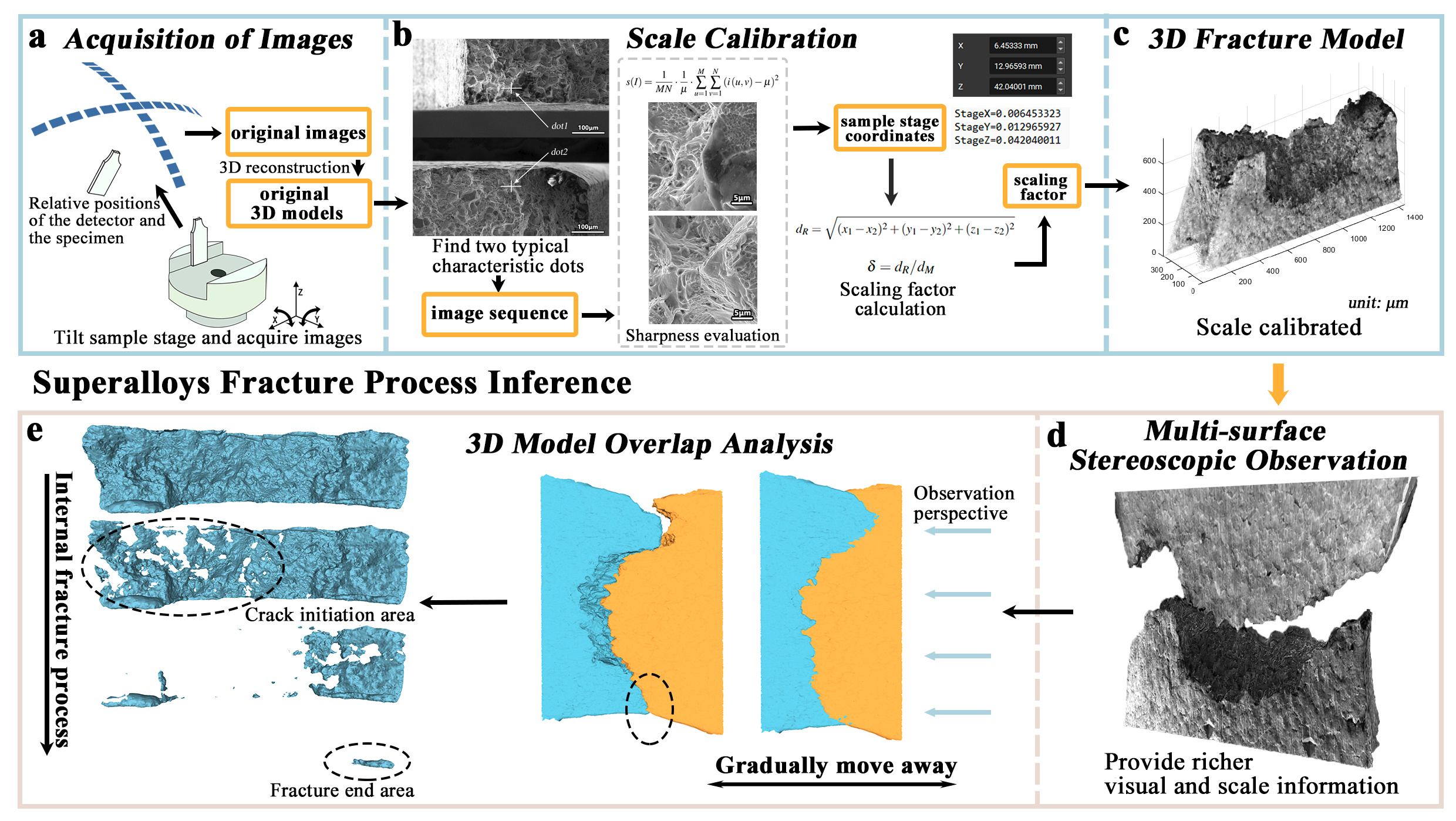
Figure 2. Overview of the proposed superalloys fracture process inference framework. a Improved image acquisition process. Images of the fracture specimens from different viewpoints were obtained by tilting the sample stage, and 3D reconstruction was performed to obtain the original 3D models. b Calculate the scaling factor to complete the 3D model scale calibration through image sharpness evaluation. c Calibrated 3D fracture morphology model in 3D coordinate system. d Multisurface stereoscopic observation. Simultaneous observation of multiple surfaces through a pair of 3D fracture morphology models to solve problems such as difficulty in matching boundary features. e The internal fracture process of the sample was obtained by inferring from the 3D model overlap analysis. The figure shows the area where the internal cracking starts and the area where the fracture ends.
To ensure the accuracy of our 3D models, we developed a scale calibration method using SEM images. This method proved to be a cost-effective and time-efficient alternative to other calibration techniques, making our approach accessible to a broader range of researchers. Figure 3 illustrates the comparison between the 3D fracture morphology model and the 2D image, as well as the results of the scale validation.
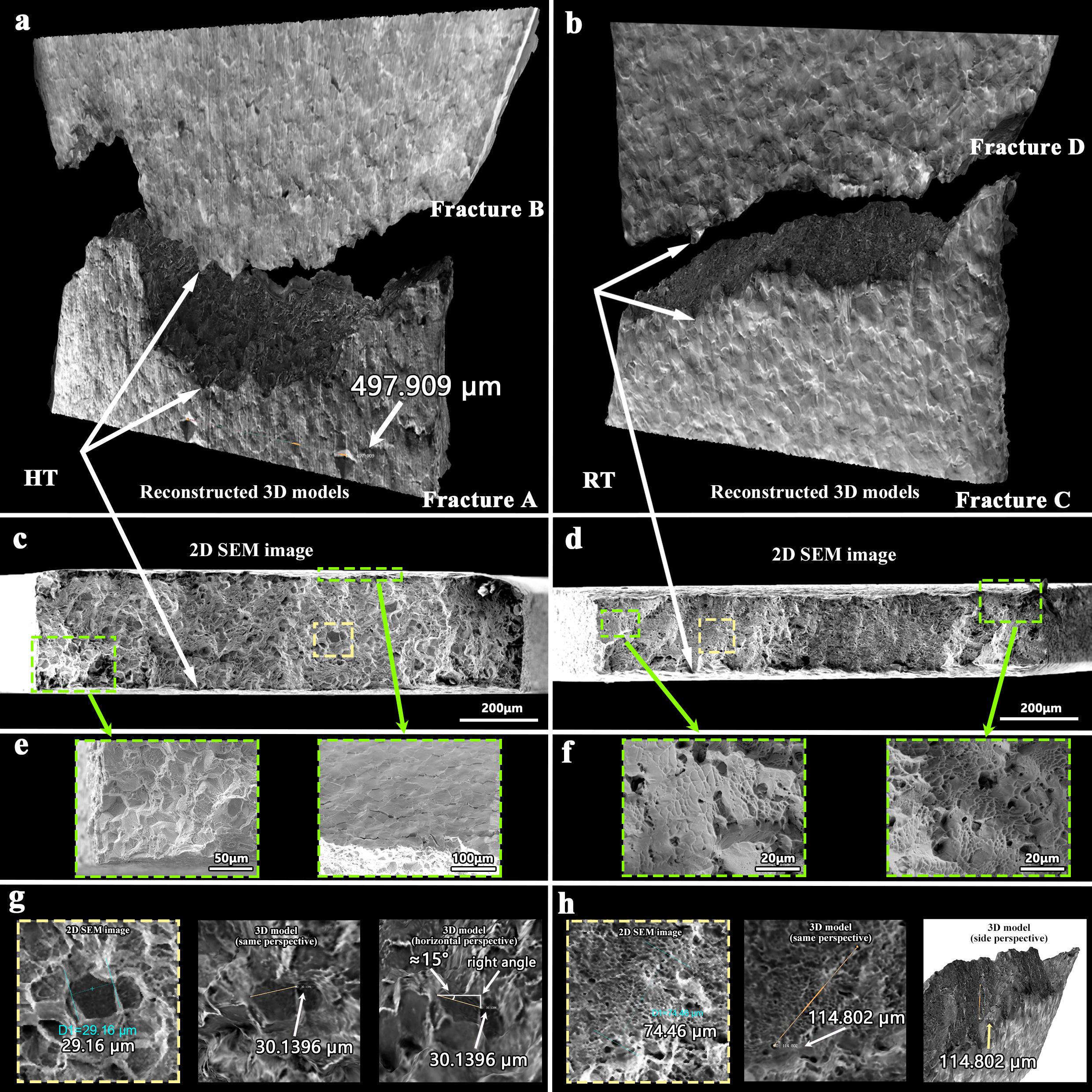
Figure 3. Comparison of 2D images and 3D stereoscopic observation and verification of scale calibration results. a, b 3D fracture surface models of IN718 after 650 ∘C high temperature (HT) and room temperature (RT) tensile experiments with an indentation spacing in the model. c, d Corresponding 2D Scanning Electron Microscope (SEM) images of the fracture surface. e Intergranular fracture at 650 ∘C. f Transgranular fracture at RT. g The distance between two diagonals of grain in an SEM image and a 3D fracture morphology model. h The distance between two void defects in an SEM image and a 3D fracture morphology model.
Challenges and Solutions
One of the significant challenges we faced was the complexity of the superalloy's microstructure, which necessitated a high level of precision in our 3D reconstructions. We overcame this by refining our image acquisition and reconstruction processes, ensuring that our models accurately represented the specimen's morphology.
Another challenge was validating our methodology. We addressed this by comparing our inferential results with in situ experimental observations, providing empirical evidence that our method could accurately predict the fracture process.
Implications and Applications
Our findings have profound implications for the design and optimization of superalloys. We can, under in-situ experimental conditions, observe the evolution of surface morphology while also inferring the internal crack propagation and evolution. By understanding the internal fracture mechanisms, engineers can tailor alloy compositions and processing methods to enhance their fracture resistance. This could lead to the development of more reliable and durable materials for critical applications in aerospace, power generation, and other high-performance industries.
Looking Forward
As we reflect on this research, we are excited about the potential of our methodology to transform how material fractures are analyzed. We envision a future where 3D fracture morphology models become a standard tool in material science, enabling researchers to predict material failure with greater accuracy.
Follow the Topic
-
Communications Engineering

A selective open access journal from Nature Portfolio publishing high-quality research, reviews and commentary in all areas of engineering.
Your space to connect: The Polarised light Hub
A new Communities’ space to connect, collaborate, and explore research on Light-Matter Interaction, Optics and Photonics, Quantum Imaging and Sensing, Microscopy, and Spectroscopy!
Continue reading announcementRelated Collections
With Collections, you can get published faster and increase your visibility.
Applications of magnetic particles in biomedical imaging, diagnostics and therapies
Publishing Model: Open Access
Deadline: May 31, 2026
Integrated Photonics for High-Speed Wireless Communication
Publishing Model: Open Access
Deadline: Mar 31, 2026


Please sign in or register for FREE
If you are a registered user on Research Communities by Springer Nature, please sign in