“When death delights in helping the living” – by unraveling a link between hepatitis E and kidney disease
Published in Biomedical Research

It has long been known that infection of the liver with the hepatitis E virus (HEV), one of the most common forms of acute liver inflammation worldwide, can be associated with kidney damage, especially in patients with impaired immune status.
However, the underlying mechanism is still poorly understood. It is conceivable that manifestations can be either due directly to replication of the virus in the infected tissues leading to cellular damage or indirectly by immunologic reactions.
The shortage of appropriate in vitro models mimicking the complexity of the human pathology and the scarcity of renal biopsies in the context of ongoing HEV infection explain the difficulties in understanding the underlying mechanisms of the pathogenesis of HEV in kidney.
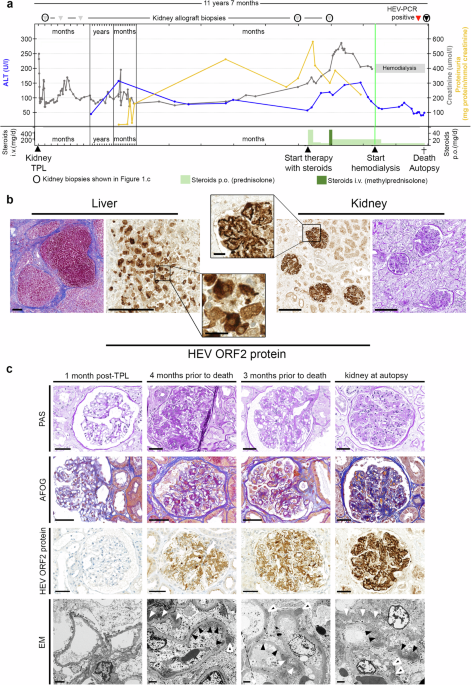
In the present study performed on HEV-infected patients with impaired immune status, we describe the journey of the main viral capsid protein, HEV ORF2 protein, starting from the replication of the virus in the liver and secretion of HEV ORF2 protein, to the formation of immune complexes in the glomeruli of the kidney.
Tissue obtained by biopsy or autopsy provides new insight
At the Department of Pathology and Molecular Pathology at the University of Zurich (UZH) and the University Hospital Zurich (USZ), we had the opportunity to better understand hepatitis E-associated kidney disease when we examined tissue from HEV-infected patients with kidney disease and impaired immune status. Through careful histopathological and molecular analysis using powerful modern microscopic and molecular biological analysis techniques, we were able to gain a deeper insight into the pathology of hepatitis E and the associated kidney damage.
Surprising detection of virus capsid protein in the kidney
Immunohistochemical tests, which we had established a few years ago for the examination of HEV infected tissues, initially pointed the way forward. The HEV ORF2 capsid protein produced in excess was detected not only in the liver but also in the kidneys, especially in the glomeruli, the filter structures of the kidneys. The retrospective examination of kidney biopsies taken in the months before the death of a patient revealed a dynamic with increasing glomerular deposition of the HEV ORF2 capsid protein in the course of chronic HEV infection and in parallel with the deterioration of kidney function. For Adriana Gaspert and Birgit Helmchen, the two renal pathology specialists in the team, from the form of the deposits, it was clear that the glomerulonephritis was caused by immune complexes and they considered the possibility that HEV was the aetiological culprit. They were able to confirm this hypothesis by means of deconvolution microscopy, in which they showed the statistically significant co-localization of the viral capsid protein and immunoglobulins G. In contrast to the liver, we found no viral RNA and therefore no evidence of viral replication in the kidney.
A hitherto undescribed form of the HEV ORF2 protein in the kidney
After it became clear that the HEV ORF2 capsid protein was the crucial molecular link between liver and kidney disease, the question arose as to which of the described variants of the HEV ORF2 protein was involved. Our initial assumption was that it corresponded to the secreted and glycosylated form of HEV ORF2 capsid protein. Surprisingly, first assessment of its molecular weight (MW) by Western Blot contradicted us as it led to a lower MW than the one expected, suggesting a truncated form of HEV ORF2 protein. At this point, we decided to get closer to the experts in the field of HEV virology, Laurence Cocquerel from the Institut Pasteur in Lille, France and Jérôme Gouttenoire and Darius Moradpour from the CHUV, Lausanne, Switzerland and refined our approach to characterize this new form. Anne-Laure Leblond and Amiskwia Pöschel patiently laser-captured all the infected glomeruli for further analysis of the protein content by mass spectrometry. Collectively, we found that a previously undescribed truncated, non-infectious and non-glycosylated form of the HEV ORF2 capsid protein accumulated in the glomeruli, where it formed immune complexes, triggering the development of glomerulonephritis.
Implications for basic research and histopathologic diagnostics
With the description of the HEV ORF2 protein-mediated immunological mechanism of kidney damage, we have not only contributed to the fundamental understanding of the development of extrahepatic manifestations in the context of hepatitis E. Our discovery also has direct implications for the histopathological diagnosis of hepatitis E-associated glomerulonephritis, as a proportion of immune complex glomerulonephritis can now be assigned to a specific etiology and reliably diagnosed using HEV ORF2 protein immunohistochemistry. We are confident that our discovery will improve the diagnosis of hepatitis E and in particular its renal involvement. We hope that our discovery will increase the awareness of hepatitis E and help to ensure that this disease is missed less frequently in future. Those affected should benefit from the unequivocal diagnosis followed by a more targeted therapy.
Reflections
Looking back on this in-depth investigation, we view our results as a combination of serendipity, teamwork and persistence. This study confirms a statement by Louis Pasteur: “…in the fields of observation, chance only favors the mind which is prepared…” as our pre-existing interest in renal involvement in HEV infection prompted additional studies on otherwise routine clinical autopsies. Eventually our scientific journey took us from the morgue to the laboratory. By use of the above mentioned methods and analysis, our study is paradigmatic for a quote often found above the entrance of old autopsy suites: “Hic locus est, ubi mors gaudet succurrere vitae” - "This is the place where death delights to help the living". In this spirit, we are deeply grateful to the patients and to their next of kin who consented to carry out and publish this work.
Follow the Topic
-
Nature Communications

An open access, multidisciplinary journal dedicated to publishing high-quality research in all areas of the biological, health, physical, chemical and Earth sciences.
Related Collections
With Collections, you can get published faster and increase your visibility.
Women's Health
Publishing Model: Hybrid
Deadline: Ongoing
Advances in neurodegenerative diseases
Publishing Model: Hybrid
Deadline: Mar 24, 2026

Please sign in or register for FREE
If you are a registered user on Research Communities by Springer Nature, please sign in