In our recent study, we unveiled the metal-oxide interfaces at the atomic scale, focusing on Zr-ZrO2 nanoparticles, using atomic-resolution electron tomography (AET). This study aimed to discover the three-dimensional (3D) atomic arrangements of metal-oxide interfaces, which are crucial for understanding the structures of natural oxidation, and encourage future materials design with metal-oxide interface. Metal-oxide interface plays an important role in numerous materials. However, due to the inherent complexity of these interfaces, especially at the atomic level, and the limitations of traditional two-dimensional (2D) imaging techniques, our understanding remains incomplete. Previous studies have largely been based on ideal atomic models or limited 2D projections, which do not reveal the true structure of these interfaces. We chose Zr and ZrO2 as our model system due to the moderate oxidation process of Zr and the strong Zr-O bonding, which provides a robust interface for tomography experiment. Our study used AET, a technique capable of visualizing atomic coordinates of Zr in 3D, to probe the Zr-ZrO2 nanoparticles. By using this advanced imaging method, we were able to overcome the resolution limitations, achieving near-atomic resolution in three dimensions.
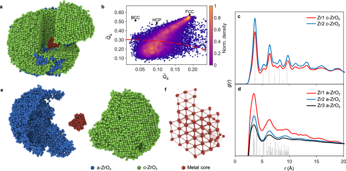
We discovered that the Zr-ZrO2 interface is not coherent; instead, it exhibits a range of structural arrangements, including semicoherent and incoherent interfaces. Atoms at these interfaces show low structural ordering and elongated bond lengths, deviating significantly from the bulk material properties. The main conclusions of our study show as following. Firstly, the 3D atomic structure revealed a complex core-shell configuration in partially oxidized Zr-ZrO2 nanoparticles, where a face-centered cubic (FCC) Zr metal crystal serves as the core, surrounded by amorphous and crystalline ZrO2. Secondly, our findings also found a variety of specific porous structures within the oxide layer, such as Zr vacancies, nano-pores, and the large pore. These voids are distributed throughout the nanoparticle and play a critical role in the oxidation process, influencing the transport of atoms and the overall stability of the interface. Finally, a gradient in oxidation was observed from the surface of the oxide shell to the metal core, with the degree of oxidation decreasing and the packing density of Zr atoms increasing toward the metal core. This gradient is important as it affects the electronic properties and reactivity of the nanoparticles.
One of the main challenges we encountered was distinguishing the positions of oxygen atoms due to their low atomic number, which provides weak contrast/intensity in annular dark-field scanning transmission electron microscopy (ADF-STEM) images. To address this, we utilized advanced structure analysis techniques and simulations to estimate oxygen contrast/intensity and positions indirectly. Additionally, the inherent disorder and non-epitaxial nature of the interfaces required careful interpretation of the data to ensure accurate representation of the atomic structure.
The results of our study have profound implications for the field of materials science, particularly in the understanding and design of metal-oxide interfaces. Our findings could pave the way for improved design strategies in various applications, including catalysts, where the activity is often governed by the atomic structure at interfaces, and in electronic devices, where metal-oxide interfaces are critical for device performance and electronic structures. What’s more, the ability to visualize these interfaces in 3D at atomic resolution provides insights into the fundamental processes governing oxidation, diffusion, and defect formation. The insights could influence the development of corrosion-resistant coatings and high-performance ceramics.
This study opens several avenues for future research. Firstly, capture the 3D atomic structure with AET in time sequence will further unveil the oxidation process. Secondly, expanding this AET technique to other metal-oxide systems could help uncover universal principles governing interface behavior. Thirdly, studying these interfaces under varying environmental conditions, such as different temperatures and reactive atmospheres, could provide further insights into the detailed and general corrosion mechanism. Moreover, integrating our experimental findings with theoretical modeling and simulations could offer a more comprehensive understanding of the energetics and kinetics involved in interface formation and evolution. This approach could help predict the behavior of similar systems under different operando conditions, enhancing the material design process.
I would like to extend my gratitude to all the authors for their contributions to this work. Special thanks to Prof. Jihan Zhou, Prof. Haibo Ke, the editor, and the reviewers for their invaluable suggestions throughout this project.
For more details, please refer to our paper “Three-dimensional atomic insights into the metal-oxide interface in Zr-ZrO2 nanoparticles” in Nature Communications (2024). (https://www.nature.com/articles/s41467-024-52026-w)
Follow the Topic
-
Nature Communications
An open access, multidisciplinary journal dedicated to publishing high-quality research in all areas of the biological, health, physical, chemical and Earth sciences.

Related Collections
With Collections, you can get published faster and increase your visibility.
Women's Health
Publishing Model: Hybrid
Deadline: Ongoing
Biosensing
Publishing Model: Hybrid
Deadline: Sep 30, 2026


Please sign in or register for FREE
If you are a registered user on Research Communities by Springer Nature, please sign in