
Tucked underneath boulder-shaped coral and nestled between the venomous spines of a sea urchin, is a fish. This fish, Siphamia tubifer, is a cryptic, unassuming species of cardinal fish. Snorkeling, you might not notice them. However, inside this fish could lie the key to gut microbiome research.
By day, Siphamia tubifer cluster around a sea urchin and sway in a coordinated dance to avoid being stabbed by one of the numerous long spines which make up their home. With the hustle and bustle of a coral reef, finding these little homebodies is almost a game of hide-and-seek. While during the day they don’t seem to do all that much, as the sun sinks, they prepare to partake in the ocean’s night life. In the shimmering moonlight, their urchin homes crawl out onto the ocean floor to feed. Siphamia tubifer are mostly black during the day, but as they drift away from the urchin, their spine-mimicking stripes melt away leaving them as pale as the sand. Camouflaged, the fish dart away to hunt for tiny organisms swimming around the reef. This is when the magic happens.
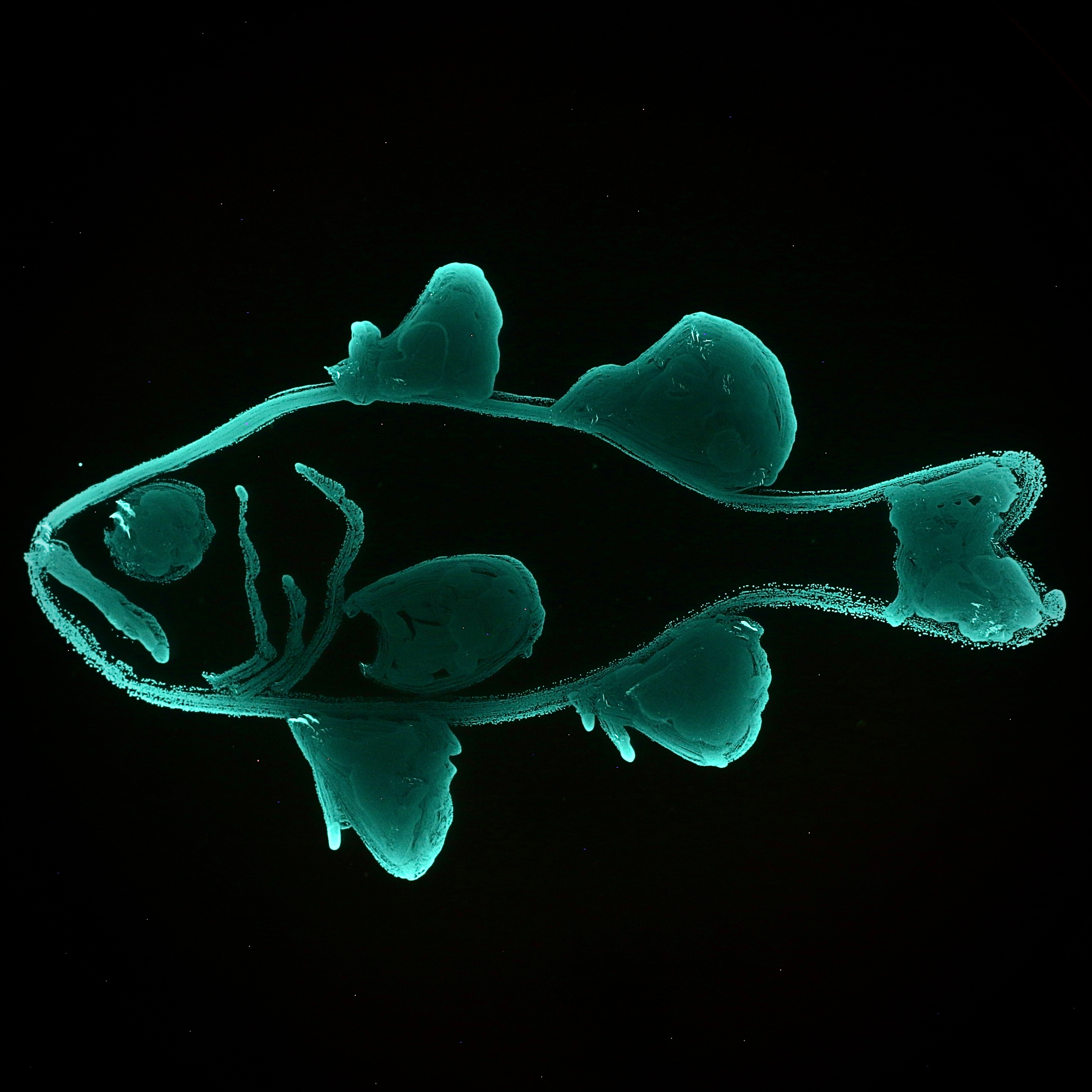
Inside each S. tubifer is a pearlescent, lentil-sized disc located around where your bellybutton is (if you were a fish, of course). This is their light organ. It is attached with a singular duct to the fish’s GI tract and is nestled within a layer of shutter-like tissue, which the fish can use to control how much light they emit. We believe this capability allows S. tubifer to counter shade against the moonlight, similar to the Hawaiian bobtail squid. Meaning, the fish can shine just enough light down onto the sea floor to cancel out their shadow, making them practically invisible to predators. Pretty handy when you are as small and appetizing as a fun-sized Twix, right? While some organisms, like fireflies, can produce their own light, many others rely on symbiotic partners to become bioluminescent. Siphamia tubifer is one of these organisms and fills their light organ with their symbiont, Photobacterium mandapamensis. The nuance between this symbiosis is where this system shines.
Defining bacterial species is an established sore spot for many microbiologists. To date, the most popular way to differentiate between bacterial species is to compare their average nucleotide identities (ANI). Those with and ANI of 95% or more are considered to be the same species. However, once you throw factors such as asexual reproduction, horizontal gene transfer, mobile genetic elements, and mutation rates it is understandable how defining these invisible, everchanging organisms is a challenge. Photobacterium mandapamensis has an ANI just above 95% to Photobacterium leiognathi, another bioluminescent bacterium. This technically makes P. mandapamensis a sub-species of P. leiognathi. However, as we are beginning to uncover, the ecological niches between the two may be different enough to define them as separate species.
Outside of its symbiosis with S. tubifer, P. mandapamensis is not habitat selective; it can live independently in the ocean and has been found on the surface of other marine animals. Other symbiotically luminous fish will associate with a range of Photobacterium species, including both P. mandapamensis and P. leiognathi. However, Siphamia tubifer will only associate with P. mandapamensis. This little, unassuming fish somehow selects for a particular sub-species of a bacterium, making this symbiosis both binary and extraordinarily specific. So, if you were to shrink yourself down into the light organ (Magic School Bus-style) you would expect to only find the same version of P. mandapamensis, right? Afterall, you just read how picky S. tubifer is in selecting its symbiont. Actually, there are multiple strains of P. mandapamensis within a light organ. In this study, we wanted to dive deeper into this strain variation within a light organ. Are there specific sets of strains found together? Do these sets vary between locations? How diverse is the symbiont pangenome? Do the strains have different characteristics from each other? What we found was a surprising juxtaposition.
Fish don’t have fingers, but these fish have a symbiont ‘fingerprint.’ Through a technique called PCR fingerprinting, we discovered light organs can contain anywhere from 1 to 13 different strains of P. mandapamensis. In fact, on average light organs contain 6 strains, and few strains are shared across individual fish. In this study we looked at fish from Japan and the Philippines, and through whole genome sequencing, we discovered that these ‘symbiont fingerprints’ seem to be region specific! Bacterial strains from both regions formed two separate clades, and we are now interested in seeing if fish from one area can accept strains from somewhere else. Furthermore, these strains can also differ significantly from each other in their growth rate and luminosity. In fact, there seems to be a tradeoff between growing fast and glowing bright. This piqued our interest, and we are now starting to unravel how these distinct strain-level symbiont communities assemble in the light organ.
These microscopic, glowing organisms which are housed comfortably in this specialized organ are just like the little ridges and dips on the fingers you’re using to scroll down this webpage right now - the same but unique. This symbiosis is extremely specific, yet the strain-level diversity is immense. Talk about a fascinating dichotomy! Now allow me to remind you that the light organ is attached to the GI tract of the fish, technically making it part of the fish’s gut. Bacterial strains and their roles in the community are often overlooked in microbiome research. This is completely understandable because most microbiomes, take our gut for example, are as simple as a box of tangled string lights. We can see the big picture, and as a field, we are just starting to unravel the complexities of microbial symbiosis. We believe the S. tubifer-P. mandapamensis system is an emerging vertebrate-bacteria model applicable to many avenues of symbiosis research. For example, what is the role of P. mandapamensis and where does it occur in the environment? How, where, and when do larval fish find their luminous bacteria? The more we think about this system, the more questions we have than answers. But to think, everything we seek is sitting inside a fish, nestled in an urchin, tucked under a coral, off a picturesque tropical beach. With just a snorkel, you could duck down into the crystal clear, warm water and find this simple fish idling away the daylight. It’s almost as if they are waiting for us. Waiting for their time to shine.


Please sign in or register for FREE
If you are a registered user on Research Communities by Springer Nature, please sign in