Continuous optical zoom microscope with extended depth of field and 3D reconstruction
Published in Physics and Protocols & Methods

Microscopes are one of the most basic types of equipment in the fields of biomedicine, micro-manufacturing, industrial inspection and other fields of science and technology. The traditional optical microscope employs discretely fixed focus objectives to change the magnification. The switching of the magnification requires manual or electronic rotation of the objective lenses, which inevitably brings about sample vibration and has a long response time. Moreover, a microscope with extended depth of field and 3D reconstruction function which can rapidly acquire clear images with different depths and generate the 3D morphology of the samples is also urgently needed.
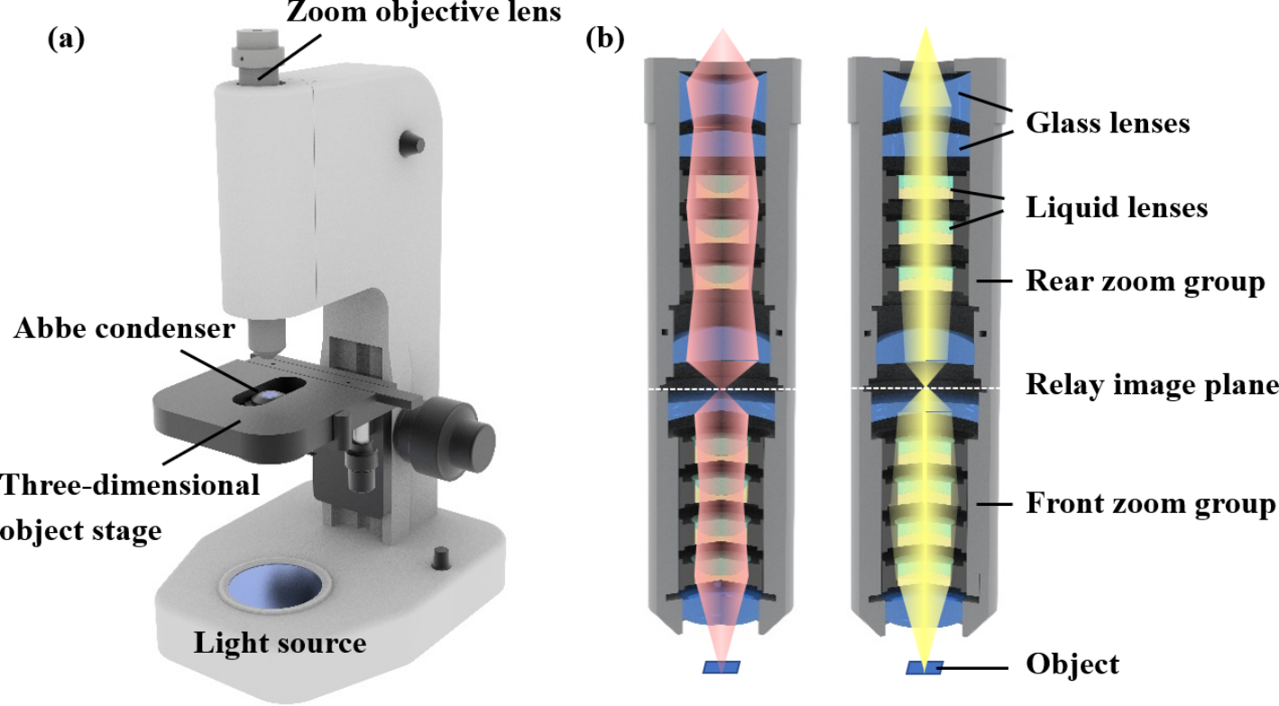
In this work, a continuous optical zoom microscope with extended depth of field and 3D reconstruction is demonstrated for the first time. The proposed microscope mainly consists of a zoom objective lens and an adjustable three-dimensional object stage. The zoom objective lens is composed of several liquid lenses and solid lenses. Figure 1 shows the schematic structure of the proposed microscope. By adjusting the voltage to the liquid lenses and using the improved image algorithm, the proposed microscope can realize large range continuous zoom imaging as well as extended depth of field. Moreover, 3D reconstruction can also be achieved without moving object stage or objective lens.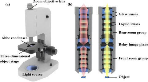
Figure 1 Schematic structure of the (a) proposed microscope and (b) the zoom objective lens.
By modulating the voltage applied to the liquid lens, the magnification of the microscope can be adaptively continuously adjusted from 10× to 60×, as shown in Figure 2. Unlike the traditional microscope, which results in sample loss due to switching magnification, the proposed microscope can always keep the observed sample in the visual field during zoom. This reduces the time delay associated with switching magnification to find the sample again. During capturing image process, the working distance does not need to be tuned, which reduces the vibration of the sample. The response time for switching magnification is measured to be around 50 ms, which is determined by our self-developed liquid lens drive board. Therefore, the proposed microscope can greatly improve the accuracy and real-time performance of microscopic observation.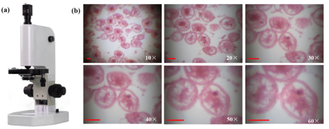
Figure 2 Proposed microscope and captured images of the sample with different magnifications.
In addition, an algorithm for Laplacian pyramid image fusion is proposed to expand the depth of field. Focused images of different depths are captured by fast adjusting the focal planes of the proposed microscope and then fused into a clear fully focused image through the proposed algorithm. On this basis, an improved focusing method algorithm based on the principle of focusing microscopy 3D reconstruction algorithm is developed, which does not require prior knowledge such as imaging models, but only performs 3D reconstruction based on the correspondence between pixel focusing degree and its depth. A value of the focus evaluation operator is designed to determine whether it has Gaussian distribution characteristics, in order to remove noise points that do not conform to Gaussian distribution. The focus evaluation operator adopts an improved sum Laplacian operator. Based on the continuity of the sample surface, the surrounding depth values are reasonably utilized to estimate the depth values at the noise points, completing the three-dimensional reconstruction of the sample. The results are shown in Figure 3.
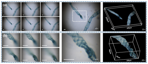
Figure 3 Depth extension and 3D reconstruction results of the proposed microscope.
To sum up, we report a continuous optical zoom microscope with extended depth of field and 3D reconstruction. By adjusting the applied voltages to the liquid lenses, the proposed microscope can achieve a large continuous magnification from ~ 10× to ~ 60× with a response time of ~ 50 ms. Under the premise of a fixed working distance, the proposed microscope can perform axial depth scanning to realize extended depth of field and 3D reconstruction. The proposed microscope can be widely applied in the fields of biological detection, chemical detection, etc.
Follow the Topic
-
PhotoniX
PhotoniX aims to present brave endeavors in promoting X-disciplinary research, latest progress of engineering applications and breakthroughs in scientific discoveries, all enabled by photonics. Original scientific letters, articles, reviews, and technology progress reports are equally welcome.

Related Collections
With Collections, you can get published faster and increase your visibility.
Photonics X Metaverse
Metaverse is a comprehensive concept because it is not a single technology, but an expansive, interconnected framework composed of multiple disciplines. Therefore, to fully realize the metaverse, it is necessary to integrate expertise from traditionally siloed fields. Photonics, among them, is acting as a powerful catalyst, driving waves of innovation across all layers of the metaverse hardware ecosystem. This special Issue aims to highlight the latest breakthroughs and insights at the intersection of metaverse and photonics and to foster continuous, cross-disciplinary dialogue and collaboration.
Scope:
Topic 1: Photonics-powered near-eye displays (magnifiers, birdbaths, waveguides, etc.)
Topic 2: Photonics-powered microdisplays (Micro LED, Micro OLED, LCoS, DMD, laser, etc.)
Topic 3: Photonics-powered three-dimensional displays (holography, light field, etc.)
Topic 4: Nanophotonics-powered lenses (freeform lenses, Fresnel lenses, pancake lenses, metalenses, contact lenses, etc.)
Topic 5: Biophotonics-powered bionics (bionic eyes, retinal implants, etc.)
Publishing Model: Open Access
Deadline: Dec 31, 2026
Solar Light-Matter Interactions: Energy Regulation, Conversion and Its Interdisciplinary Applications
Scope:
Topic 1: Fundamental physical and chemical mechanisms underlying solar light-matter interactions
Topic 2: Design, fabrication, and solar radiation regulation performance of micro/nanostructures (e.g., metamaterials, plasmonic materials, porous materials)
Topic 3: High-efficiency solar photothermal/photovoltaic conversion devices and systems
Topic 4: Daytime radiative cooling materials, devices, and their engineering applications
Topic 5: Applications of solar energy conversion technologies in water treatment, environmental remediation, building energy efficiency, and other related fields
Topic 6: Research on the photothermal dynamic regulation mechanisms between solar light and organisms (including humans and other mammals, etc.)
Publishing Model: Open Access
Deadline: Ongoing
Please sign in or register for FREE
If you are a registered user on Research Communities by Springer Nature, please sign in