Human connective tissue
Published in Anatomy & Physiology
Behind the science
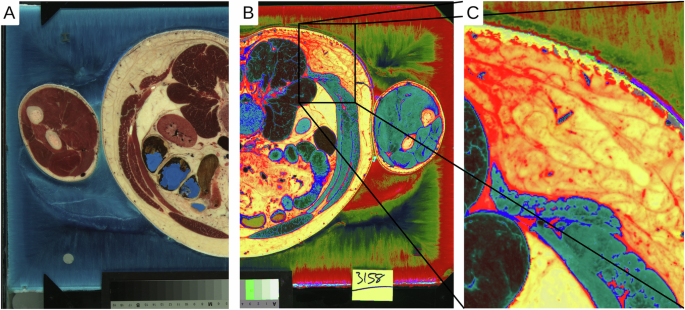
Some studies are planned, while others arise from problems. This study falls into the latter category. It began with the question of how to easily reconstruct the musculature from the data of the Visible Human Project. The project focused on the prevention of back pain (Movies). The bones could be segmented quite easily from the corresponding CT data set. However, it was more difficult to segment the musculature in the scanned colour images of the cross-sections. Although the musculature can be identified quite clearly by its red to brown tones, but there are a number of organs that have a similar colour tone (lungs, liver, and kidneys). The first thing to try is to separate the different shades of red from each other. This can be done quite easily in an image editing software (e.g. Gimp) by remixing the colours and adjusting their brightness. Afterwards, certain colour areas can be separated. When separating the muscles, it became apparent that the connective tissue could also be separated in this way.

What next?
This information was initially deposited on the shelf for a while, as the main focus of research was on the musculature. Over the years, however, it became increasingly clear that connective tissue plays a very important role within the musculature. It plays a key role in transferring forces and holding everything together. Furthermore, this is not limited to the musculature, but affects the entire body. The first step was to segment the connective tissue individually. After that, the next step was to identify individual morphological parameters. This was based on biomechanical parameters. In other words, how is the connective tissue organised (fibrous, planar) and what is the local proportion? This can be analysed using suitable methods. The final step was to make it available to all scientists.
Structure and function on micro- and macroscopic levels for cells/tissues/species related to metabolism, locomotion and immune system.
Follow the Topic
-
Scientific Data
A peer-reviewed, open-access journal for descriptions of datasets, and research that advances the sharing and reuse of scientific data.

Related Collections
With Collections, you can get published faster and increase your visibility.
Computer vision in plant science and agriculture
Publishing Model: Open Access
Deadline: Oct 10, 2026
Wearable and Computer Vision Data for Health and Behaviour Research
Publishing Model: Open Access
Deadline: Aug 08, 2026


Please sign in or register for FREE
If you are a registered user on Research Communities by Springer Nature, please sign in