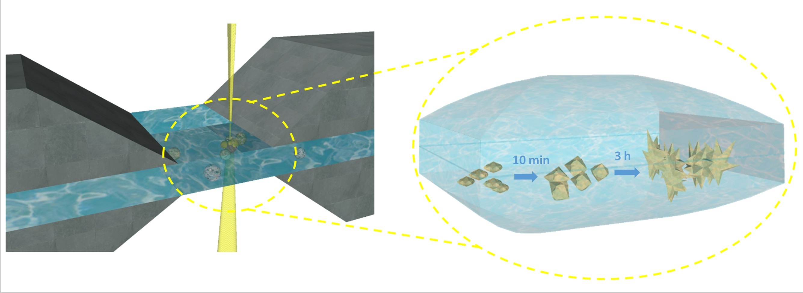
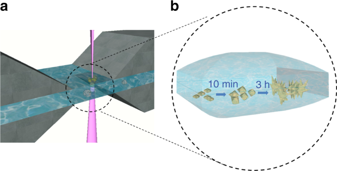
It all started when we got together with my colleague Kathryn Grandfield and we talked about how important it would be to apply liquid cell transmission electron microscopy (TEM) to study relevant biological systems. Calcium phosphate biomineralization immediately came to mind. This is an essential process in the formation of bones and teeth. It is important in pathological calcification like the formation of kidney stones, and knowing more about the process can guide us in engineering bio-inspired materials.
In the work published in Communications Chemistry, we used liquid cell TEM to study calcium phosphate mineralization from stimulated body fluid. Under low-beam imaging conditions, we were able to create a nano-movie that showed the structural evolution of calcium phosphate, directly in liquid, with nanoscale spatial resolution and sufficient temporal resolution. The nano-movie demonstrated that calcium phosphate mineralizes through a non-classical pathway, by the formation of pre-nucleation particles and aggregative growth forming chain-like or branched morphologies. This study demonstrates the feasibility of studying biological processes in their native liquid environment with nanoscale resolution using liquid-cell TEM
Follow the Topic
-
Communications Chemistry
An open access journal from Nature Portfolio publishing high-quality research, reviews and commentary in all areas of the chemical sciences.

Related Collections
With Collections, you can get published faster and increase your visibility.
Chemical modification of proteins
Publishing Model: Open Access
Deadline: Sep 30, 2026
Sustainable waste management through polymer upcycling
Publishing Model: Open Access
Deadline: Aug 31, 2026


Please sign in or register for FREE
If you are a registered user on Research Communities by Springer Nature, please sign in