Single proton counting using transmissive perovskite nanocrystal scintillators
The use of high-energy ion beams, particularly proton beams in cancer radiotherapy has expanded rapidly in recent decades. This method is preferred over conventional X-ray and γ-ray therapy as it enables maximum dose delivery directly to the tumor, minimizing exposure to surrounding healthy tissues. To ensure precise dosage in proton therapy, accurate dose measurement is crucial. Current dosimeters such as ionization chambers, silicon-based detectors, and plastic scintillators aim to quantify protons accurately and in real time, ideally at the single proton level.
However, there are challenges. Ionization chambers and silicon detectors are too bulky, hindering their ability to deliver protons to the patient in real time. Plastic scintillators, particularly in the form of scintillating fibers, are invasive and have limitations in detection accuracy and radiation durability. Therefore, the development of noninvasive, effective, and real-time detectors for accurate proton counting remains a challenge. Current alternatives, such as thin-film diamond detectors and organic scintillators, face their own issues. Thin-film diamonds are complex and costly to fabricate, while organic scintillators yield lower scintillation due to their low atomic electron density. This has led to the exploration of high- and high-density materials for scintillators. In this context, transmissive thin scintillators made from CsPbBr3 nanocrystals have emerged as a promising solution for efficient proton detection.
Proton-induced luminescence and mechanism
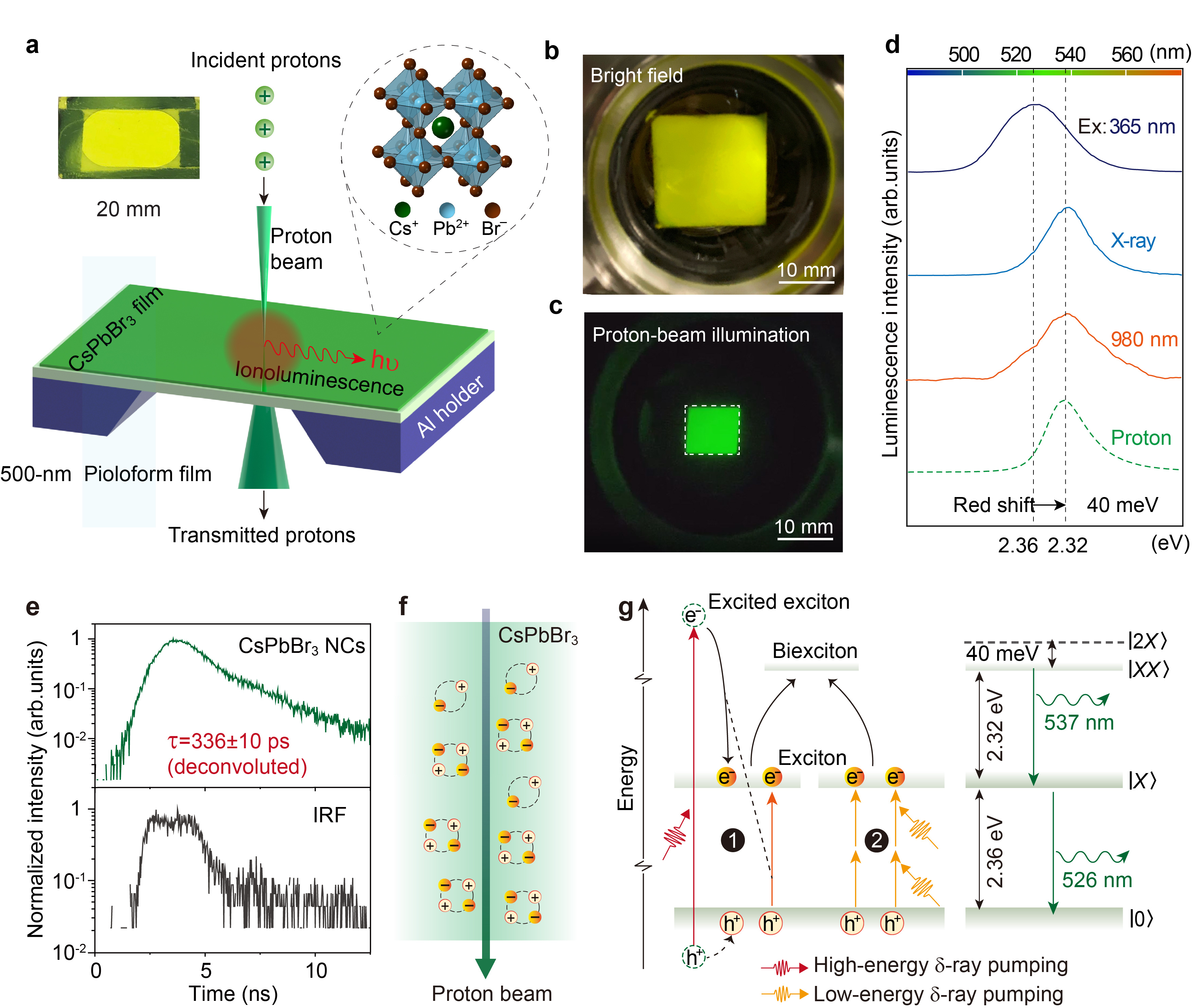
For our study, we utilized an ion accelerator equipped with a hydrogen source to generate a proton beam with a typical kinetic energy of 2 MeV. When this proton beam irradiated the CsPbBr3 nanocrystals film, we observed a strong ionoluminescence effect. The ionoluminescence spectrum was centered at 537 nm, corresponding to an energy of 2.32 eV. Notably, when compared to the emission at 365 nm excitation, the proton-induced emission exhibited a red shift and a narrower full width at half maximum (FWHM), similar to the pattern observed under X-ray and near-infrared (~980 nm) excitation.
We next investigated the underlying mechanisms of this scintillating ionoluminescence. We first performed theoretical calculations to determine the frequency of production of d-rays in CsPbBr3 nanocrystals. These calculations were based on the Hansen-Kohach-Stolterfoht model. Our findings indicated that the kinetic energy of these d-rays ranged from sub-eV to about 4.3 keV, and that their production frequency decreased rapidly with increasing energy. We further substantiated our theoretical predictions by experimentally measuring the back-emitted electrons from the surface of a CsPbBr3 scintillator sample.
In our exploration of the origins behind the red-shifts in luminescence emission, we measured the emission lifetimes of CsPbBr3 nanocrystal scintillators under different excitation sources. The results indicated that the decay time scale under UV and NIR light excitation was consistent, on the order of 10 ns. This duration aligns with the previously reported lifetime of CsPbBr3 nanocrystals upon X-ray excitation. However, a strikingly different result emerged under proton-beam excitation. The emission lifetime of the CsPbBr3 nanocrystal scintillator in this context was markedly shorter, around ~336 ps. This fast decay is characteristic of multiexcitons in nanocrystals, likely due to an efficient Auger process. Since re-absorption does not alter the emission lifetime significantly, we speculate that the red-shift observed under proton-beam excitation results from the formation of multiexcitons, specifically biexcitons. These biexcitons are complex four-body quasiparticles that arise from the interaction of two excitons.

Proton-induced luminescence from CsPbBr3 nanocrystal scintillators. a, Schematic of proton-beam-induced luminescence (ionoluminescence) in a thin transmission scintillator comprising CsPbBr3 nanocrystals. b-c, Experimental demonstration of proton-induced scintillation from a CsPbBr3 nanocrystal scintillator. (b, bright-field imaging of the scintillator; c, proton-beam illumination within the square marked in b) d, Luminescence emission spectra of CsPbBr3 nanocrystals under excitation with a 365-nm light source, 40-keV X-rays (99 μA), a 980-nm laser, and 2-MeV protons, respectively. e, Time-resolved ionoluminescence measurement. f, Schematic of biexcitons as four-body quasiparticles. g, Proposed mechanism of proton scintillation in a CsPbBr3 nanocrystal.
Single-proton counting
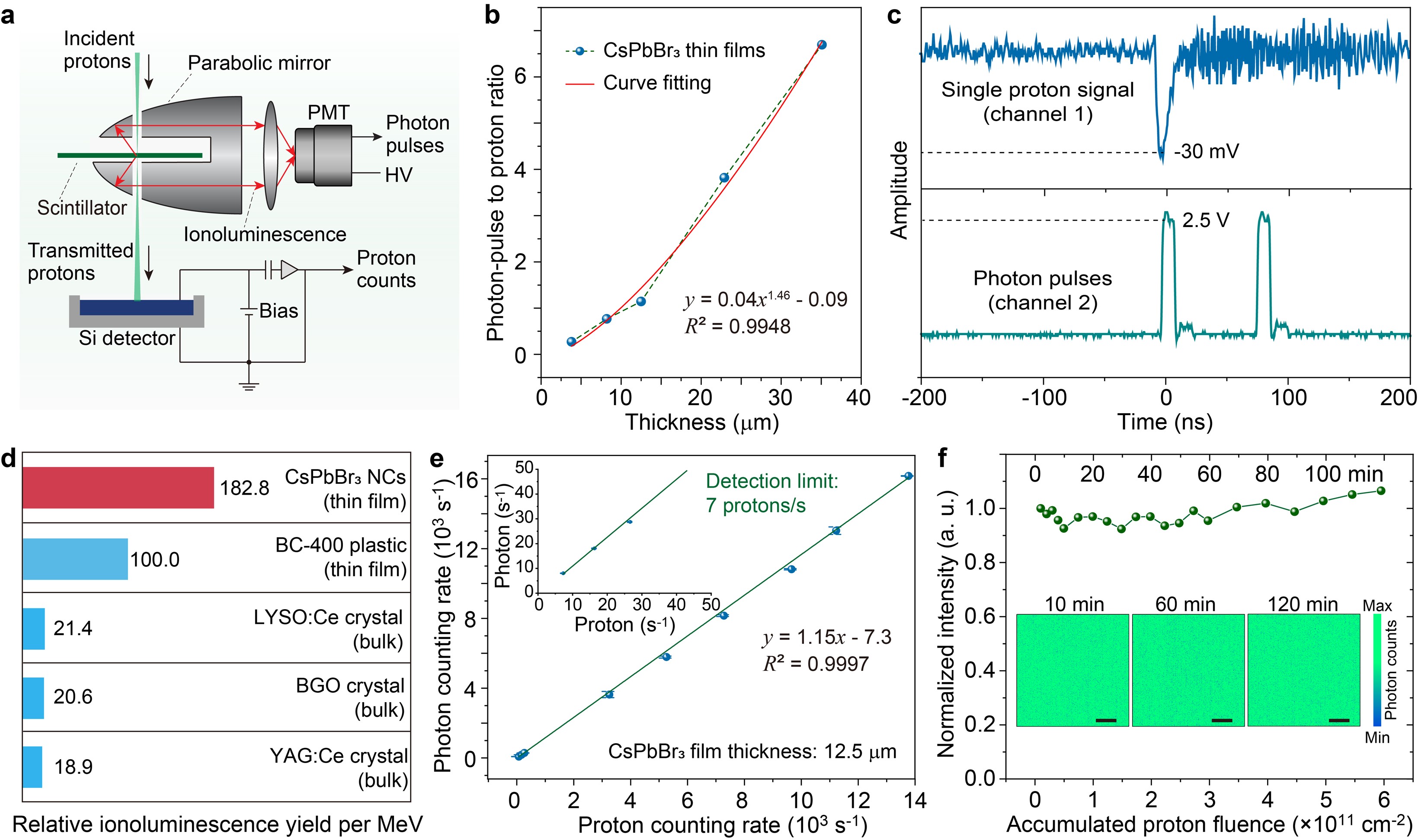
The fast response of CsPbBr3 nanoscintillators to proton beams, approximately 336 ps, is crucial advantage, particularly in scenarios where a vast number of protons are encountered in a short time. This quick response ensures that the counting of protons does not overlap, even in high-density proton environments. Consequently, CsPbBr3 nanocrystal scintillators demonstrate the necessary speed to count individual protons accurately. To confirm the capability of CsPbBr3 nanocrystal thin films in single-proton counting, we conducted a direct experiment. We traced the signals of a single proton and the resultant proton pulses using a fast oscilloscope. In this case, only one proton was present within a 400-nanosecond time domain, and the proton counting rate was maintained at a sufficiently low level of ~500 protons per second.
The CsPbBr3 nanocrystal thin film exhibited a superior relative ionoluminescence yield compared to some commercial proton scintillators. This enhanced sensitivity to proton scintillation is particularly beneficial for detecting very low levels of proton flux, with the minimum detectable rate being as low as 7 protons per second. Moreover, CsPbBr3 scintillators exhibited superior stability in photon emission when subjected to continuous proton irradiation.

Performance characterization and single-proton counting with CsPbBr3 nanocrystals. a, Schematic of the experimental set-up for single-proton counting and calibration. b, Quantum yield of CsPbBr3 nanocrystal scintillators, characterized by the ratio of photon-pulses to proton counts, as a function of scintillator thickness. c, Single proton tracing in perovskite nanocrystal thin films (thickness of ~15 µm). d, Relative ionoluminescence light yield per MeV of a CsPbBr3 nanocrystal thin-film scintillator compared with commercial scintillators under excitation by a 2-MeV proton beam. e, Measurement of ionoluminescence photon counting rate as a function of proton counting rate with a 12.5-µm-thick CsPbBr3 scintillator. f, Ionoluminescence intensity profile as a function of accumulated proton fluence.
Single-proton irradiation and high-resolution imaging
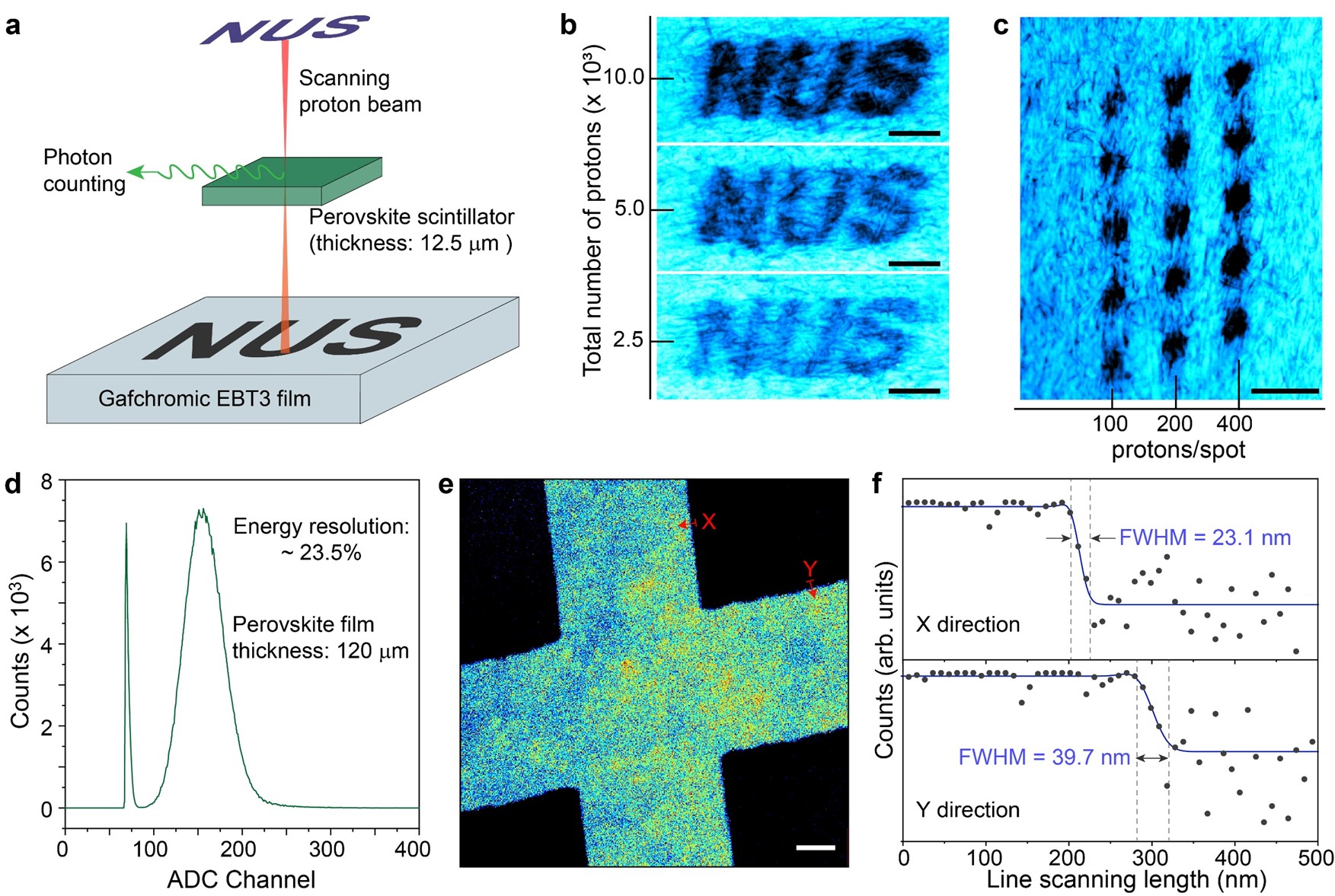
Incorporating the real-time single-proton irradiation capabilities of CsPbBr3 nanocrystal films, we proceeded to use a Gafchromic EBT3 film to capture the dose from accumulated single protons. This approach allowed us to create a series of visually distinguishable letters “NUS” on the film. These letters, appearing in varying shades from light to dark, corresponded to different numbers of implanted protons, and were clearly identifiable through optical imaging.
Our investigation then extended to accessing the potential of CsPbBr3 nanocrystals scintillators for high-resolution proton imaging. In this evaluation, we measured an energy resolution of 23.5% based on the FWHM of the spectrum. The lateral resolutions of the images were 23.1 nm horizontally and 39.7 nm vertically, suggesting that CsPbBr3 nanocrystals are excellent candidates for high-resolution proton imaging.

Single-proton irradiation and high-resolution proton imaging using CsPbBr3 nanocrystal scintillators. a, Schematic of simultaneous single-proton counting and irradiation on a Gafchromic EBT3 film. b, Optical imaging of the Gafchromic EBT3 film irradiated with the “NUS” pattern. c, Optical imaging of the Gafchromic EBT3 film irradiated with a spot array. d, Energy-resolved spectrum of 2.1 MeV protons detected with a 120-μm-thick CsPbBr3 scintillator coupled to a photomultiplier tube. e, Image showing two crossed bars of a nickel grid, taken by measuring the energy loss of protons as they penetrate the grid pixel by pixel using the CsPbBr3 scintillator. f, Cross-sectional line profiles extracted along the arrows in the X and Y directions shown in e.
In summary, our research has demonstrated CsPbBr3 nanocrystals as a new class of scintillators, ideally suitable for the high-sensitivity detection and counting of single accelerated protons. Our experimental and theoretical findings suggest that the scintillation caused by protons is primarily due to the population of the biexcitonic state in these nanocrystals. Accordingly, CsPbBr3 nanocrystals demonstrate fast and stable ionoluminescence emissions, with a much higher light yield than commercial proton scintillators. These unique characteristics of CsPbBr3 nanocrystals make it possible to develop ultrathin and flexible perovskite scintillators, which are highly adept for real-time ultrasensitive single-proton counting. We believe that CsPbBr3 nanocrystal films represent a promising option for single-proton level detection. Looking forward, while acknowledging the challenges ahead, we are optimistic about the immense potential these nanocrystals hold for proton therapy and radiography, offering significant improvements in both fields.
Follow the Topic
-
Nature Materials

A monthly multi-disciplinary journal that brings together cutting-edge research across the entire spectrum of materials science and engineering, including applied and fundamental aspects of the synthesis/processing, structure/composition, properties and performance of materials.
Your space to connect: The Polarised light Hub
A new Communities’ space to connect, collaborate, and explore research on Light-Matter Interaction, Optics and Photonics, Quantum Imaging and Sensing, Microscopy, and Spectroscopy!
Continue reading announcement


Please sign in or register for FREE
If you are a registered user on Research Communities by Springer Nature, please sign in