Two-photon fluorescence imaging and specifically biosensing of norepinephrine on a 100-ms timescale
Published in Chemistry

Nerve signal transduction in the brain includes electrical signal transduction and chemical signal transduction, the latter of which depends on the release, transmission and combination of neurotransmitters between synapses, and its time scale is as low as milliseconds. Therefore, monitoring the dynamic changes of neurotransmitters at the molecular scale is crucial for understanding the structure and function of neurons and the brain. Among them, norepinephrine (NE) is considered as one of the key neurotransmitters in the central nervous system of vertebrate organisms, and the dysfunction of noradrenergic transmission is closely related to a series of neurodegenerative and psychiatric disorders, including Parkinson’s disease (PD), Alzheimer’s disease (AD), depression, etc. Despite its well-recognized importance in a variety of physiological and pathophysiological processes, monitoring the transient NE dynamics in living systems with high sensitivity and high spatiotemporal resolution remains a formidable challenge.
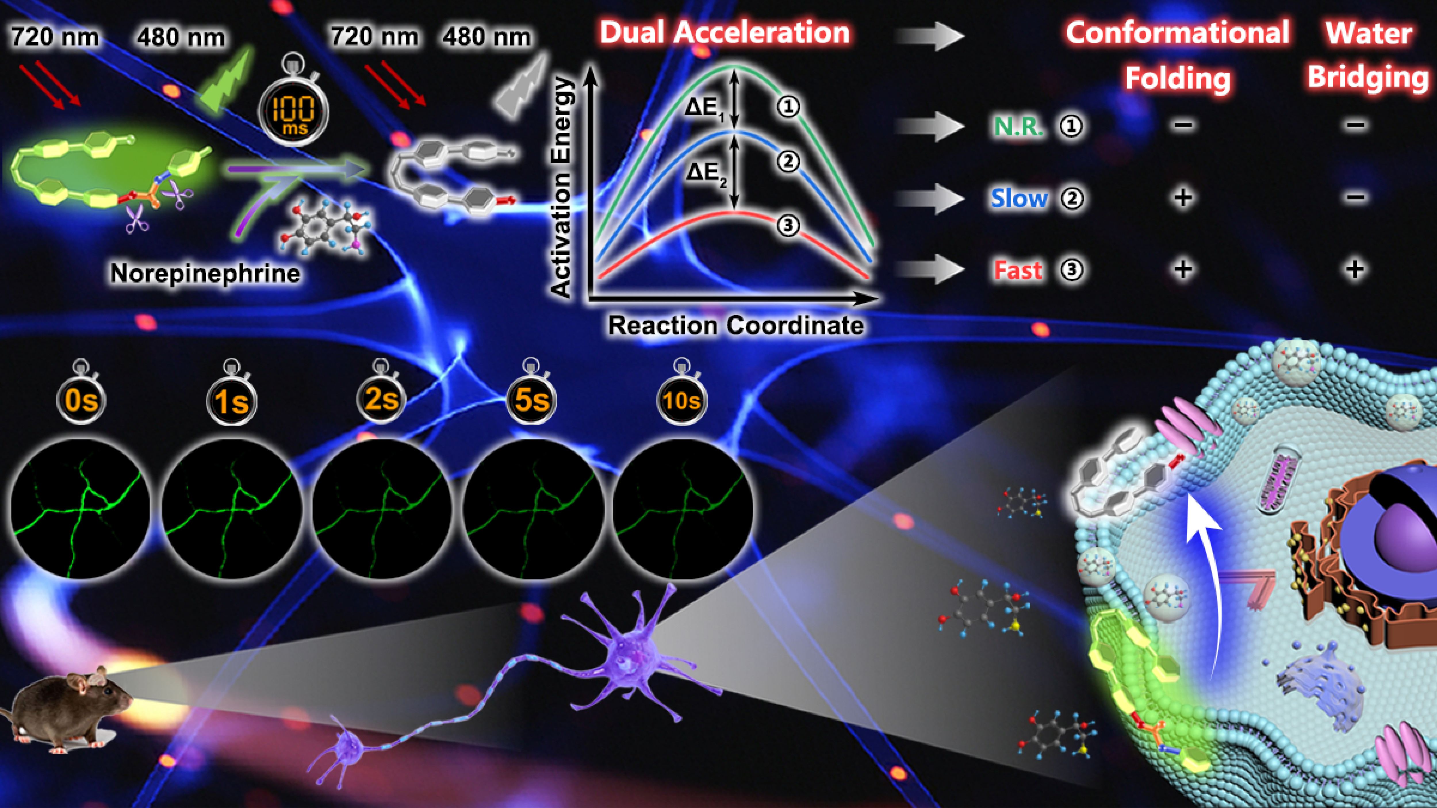
Our group recently designed and synthesized a novel small molecular fluorescent probe (BPS3, Figure 1), bearing two phenyl pyridiniums linked by an alkyl chain, which not only guaranteed good water solubility and a flexible molecular conformation, but also exhibited excellent cytomembrane targeting. In addition, the introduction of a terminal S-p-toluene carbonothioate as the triggering group for NE, which could undergo a sequential nucleophilic substitution followed by a cyclization reaction to the characteristic primary amino group and β-hydroxyl group of NE, respectively, achieved excellent selectivity toward NE over the other two catecholamine neurotransmitters (dopamine and epinephrine). The most noteworthy feature of the developed probe BPS3 is its ultrafast reaction kinetics, enabling NE detection within an impressively short time down to 100 ms, which is the most rapid small molecular probe reported by far for quantitative NE sensing and imaging. The fluorescence intensity of the probe has a good linear relationship with NE in the concentration range of 0-200 nM, and the detection limit is as low as 0.5 nM, which meets the linear range and sensitivity requirements for the detection of low concentration NE in neurons.

Through experiments and theoretical analysis, we revealed a unique dual acceleration mechanism, namely molecular conformational folding and water bridging. Through the study of molecular conformational regulation, it is confirmed that the reaction rate of the probe is more than 105 times higher in the folded conformation than in the stretched conformation (Figure 2a, b). On the other hand, through the analysis of the reaction transition states, it is proved that water molecules participate in the formation of a six-membered-ring transition state, which promotes the process of intermolecular proton transfer, thereby effectively reducing the reaction energy barrier. Experimental results demonstrate that the reaction rate of the probe is 103 times higher in the water environment than that in the anhydrous environment (Figure 2c, d).

Finally, benefiting from the superior properties including high specificity, nanomolar sensitivity, neuronal cytomembrane-labelling and 100-ms temporal resolution, as well as near infrared (NIR) two-photon excitation, we applied probe BPS3 to real-time imaging of NE fluctuations in living neurons under external chemical stimulation. The rapid and significant fluorescence variations at the neuronal cytomembrane clearly indicated the transient neuronal NE cytosolic process (within 2 s) upon high potassium stimulation, validating the high spatiotemporal resolution of the probe, which was very beneficial for in-depth studies of the transient NE dynamics and functions during physiological processes in living neurons. Moreover, by two-photon fluorescence imaging of acute mouse brain tissue slices, we revealed the close correlations between downregulated NE levels and AD pathology as well as antioxidant therapy.

The results presented here not only provided a superior molecular imaging tool for NE detection that is beneficial for advanced neuroscience research, but also revealed a dual acceleration mechanism that may further bring new inspiration to biological, analytical, organic, and physical chemists in the study of ultrafast detections and reaction kinetics regulations.
For more detailed information, see our article " Two-photon fluorescence imaging and specifically biosensing of norepinephrine on a 100-ms timescale" in Nature Communications (https://www.nature.com/articles/s41467-023-36869-3).
The brief introductions of the corresponding authors (Prof. Dr. Qi-Wei Zhang and Yang Tian) are as follows.
Prof. Qi-Wei Zhang
Qi-Wei Zhang, obtained his Ph.D. from East China University of Science and Technology in 2014 under the supervision of Prof. Tian He, an academician of Chinese Academy of Sciences. From 2014 to 2018, he successively conducted postdoctoral research at Radboud University in the Netherlands and the University of Notre Dame in the United States. The co-advisors were Prof. Roeland J.M. Nolte and Prof. Bradley D. Smith. In Sep-2018, he joined East China Normal University as a professor of Chemistry. Prof Zhang mainly engaged in the research of small molecular and supramolecular optical functional materials, fluorescent probes and biosensing, etc. He has published 29 SCI papers in journals such as Chem. Rev., Nat. Commun., JACS, Angew. Chem., Adv. Sci., Chem. Sci., Org. Lett., Anal. Chem., ACS Sens., etc. which have been cited more than 1,900 times. He has also been invited to contribute an academic book chapter named "Near-Infrared Organic Materials for Biological Applications" (CRC Press, Taylor & Francis Group, 2022).
https://orcid.org/0000-0003-1527-0559
https://faculty.ecnu.edu.cn/_s34/zqw_en/main.psp
Prof. Yang Tian
Professor Yang Tian is a Distinguished Professor of East China Normal University, and is currently the dean of School of Chemistry and Molecular Engineering. Professor Tian's team has been engaged in the analysis and detection of in vivo electrical and chemical signals for a long time. They have developed precise analysis and measurement strategies for brain neurochemical molecules, established long-term stable high-spatial resolution brain imaging methods, and developed new high-speed imaging analysis instruments. So far, she has published more than 160 papers, including Acc. Chem. Res., Nat. Commun., Sci. Adv., JACS, Angew. Chem., Adv. Mater., Adv. Sci., Anal. Chem., etc., which have been cited more than 11,000 times. She was selected as one of the ESI Highly Cited Scholars. She has been authorized 8 Chinese invention patents, are contributed to a series of book chapters on neurochemical analysis and fluorescent bioimaging. She received the funding of National Fund for Distinguished Young Scholars, and has been announced as a Chief Scientist of the Key Research and Development Program of the Ministry of Science and Technology, the Female Analytical Chemist of the Chinese Chemical Society. She won the first prize of the China Association for Instrumental Analysis, the first prize of the Shanghai Natural Science Award, etc., and was selected into the National Ten Thousand Talents Program for scientific and technological innovation Leading talent. Currently, she is the associate editor of the journal Chemical Communications.
Qi-Wei Zhang, obtained his Ph.D. from East China University of Science and Technology in 2014 under the supervision of Prof. Tian He, an academician of Chinese Academy of Sciences. From 2014 to 2018, he successively conducted postdoctoral research at Radboud University in the Netherlands and the University of Notre Dame in the United States. The co-advisors were Prof. Roeland J.M. Nolte and Prof. Bradley D. Smith. In Sep-2018, he joined East China Normal University as a professor of Chemistry. Prof Zhang mainly engaged in the research of small molecular and supramolecular optical functional materials, fluorescent probes and biosensing, etc. He has published 28 SCI papers in journals such as Chem. Rev., Nat. Commun., JACS, Angew. Chem., Adv. Sci., Chem. Sci., Org. Lett., Anal. Chem., ACS Sens., etc. which have been cited more than 1,900 times. He has also been invited to contribute an academic book chapter named "Near-Infrared Organic Materials for Biological Applications" (CRC Press, Taylor & Francis Group, 2022).
Follow the Topic
-
Nature Communications
An open access, multidisciplinary journal dedicated to publishing high-quality research in all areas of the biological, health, physical, chemical and Earth sciences.

Related Collections
With Collections, you can get published faster and increase your visibility.
Women's Health
Publishing Model: Hybrid
Deadline: Ongoing
Tumor Microenvironment Crosstalk and Therapeutic Implications
Publishing Model: Hybrid
Deadline: Nov 02, 2026


Please sign in or register for FREE
If you are a registered user on Research Communities by Springer Nature, please sign in