Unlocking the power of investigative biopsies in glioma
Published in Cancer, Biomedical Research, and Surgery
Glioblastoma is the most common primary brain cancer in adults and follows an aggressive course despite surgical resection, chemotherapy and radiation. Numerous clinical trials have been conducted and overall results have been disappointing. Trial endpoints are often crude measures of efficacy, and it is often not clear how or why the trial failed. Ideally, we could have a "window" into the tumor microenvironment during the course of treatment in order to gain a better understanding into tumor response.
Teaching an old dog new tricks
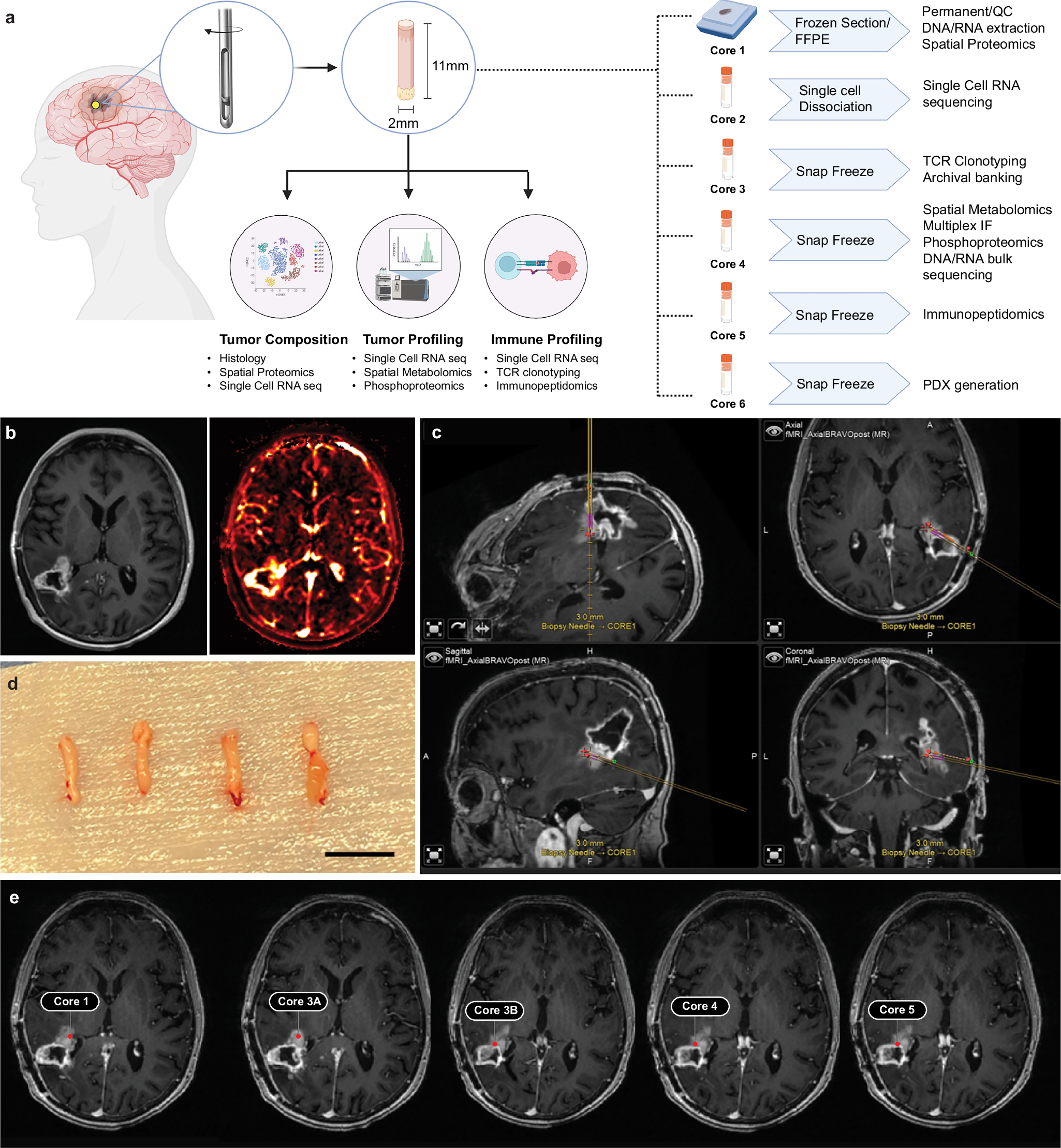
Stereotactic needle core biopsies are part of the routine neurosurgical armament, yet its use is currently restricted to diagnosis alone. The procedure involves an incision and burr hole through the skull, followed by passage of a needle to the target lesion for sampling. Stereotaxy is used to accurately guide the needle, and the amount of material is small (<50mg), but sufficient for histological and basic molecular diagnosis.
The central premise of the current study was to introduce the concept of an investigative biopsy to gain as much information as possible about the tumor state under treatment. Which naturally led to the question: Can we do more on these small samples? Can we take advantage of current multi-omics platforms to maximize the amount of information captured?
Leveraging Team Science
The impetus for this research was the assembly of a GBM "Team Lab" via the Break Through Cancer Foundation. This was a collaborative effort bringing in researchers from 5 separate institutions to form a cohesive cross-institutional research group.
We were interested in understanding tumor evolution under therapy and this became the organizing principle when selecting multi-omics assays—central to this was the generation of single cell RNA sequencing data from these samples, in combination with spatial transcriptomics, metabolomics, proteomics, and immunopeptidomics to provide a comprehensive overview of the tumor at a specific moment in time.
While the ideal and eventual goal was to sample the tumor under therapy over multiple timepoints, we first needed to determine whether the input material was sufficient for informative downstream multi-omics analysis. There was considerable skepticism at the beginning that any useful data could be generated from such small samples. Then there was the logistical aspect itself. Even if it were possible to do these analyses, the practical challenges of transporting fresh and frozen samples to different sites across the country had to be overcome if this approach was to be expanded into a clinical trial setting.
This is where the power of team science came into its own, through weekly meetings, protocol sharing and trial and error we progressively settled on a methodology we thought would work in a trial setting with the ultimate goal of performing serial biopsy sampling in GBM patients undergoing therapy. The challenge then became to demonstrate that we could do this in a real world setting to prove the feasibility of this approach.
Integrating Data Modalities and understanding Tumor Heterogeneity
Once we were able to generate the data, we next faced the task of integrating various modalities. The success of the single cell RNA sequencing provided an axis through which we could integrate different modalities and we also took advantage of orthogonal assays to cross-validate some of the existing inference algorithms.
Now that we knew we could generate high quality datasets from biopsies, one of the questions that came up in our continuing group discussions was how to resolve or account for tumor heterogeneity. We therefore devised a system for measuring tumor heterogeneity based on histological features so that we could use basic histological information to account for heterogeneity between core biopsies when integrating the multi-omics datasets. As an additional study we also looked into whether biopsies can be used to generate tumor avatars (spoiler alert: they can).
This was truly a tremendous team effort from the Accelerating GBM Therapies Through Serial Biopsies Team Lab, and the future of GBM therapies relies on us gaining a better understanding of the mechanisms of treatment response and failure. The re-purposing of routine stereotactic biopsies for advanced multi-omics analysis affords researchers an unprecedented window into the tumor microenvironment under treatment, unlocking the power of investigative biopsies.
Follow the Topic
-
Nature Communications
An open access, multidisciplinary journal dedicated to publishing high-quality research in all areas of the biological, health, physical, chemical and Earth sciences.

Ask the Editor – Inflammation, Metastasis, Cancer Microenvironment and Tumour Immunology
Got a question for the editor about inflammation, metastasis, or tumour immunology? Ask it here!
Continue reading announcementRelated Collections
With Collections, you can get published faster and increase your visibility.
Women's Health
Publishing Model: Hybrid
Deadline: Ongoing
Biosensing
Publishing Model: Hybrid
Deadline: Sep 30, 2026


Please sign in or register for FREE
If you are a registered user on Research Communities by Springer Nature, please sign in