Make the invisible visible in now possible: NIR-HSI, Bone collagen, and Radiocarbon dating
Published in Chemistry

It sounds obvious how rare and precious human remains found by archaeologists are, in particular, if we think about human bones or bone artefacts from Prehistory.
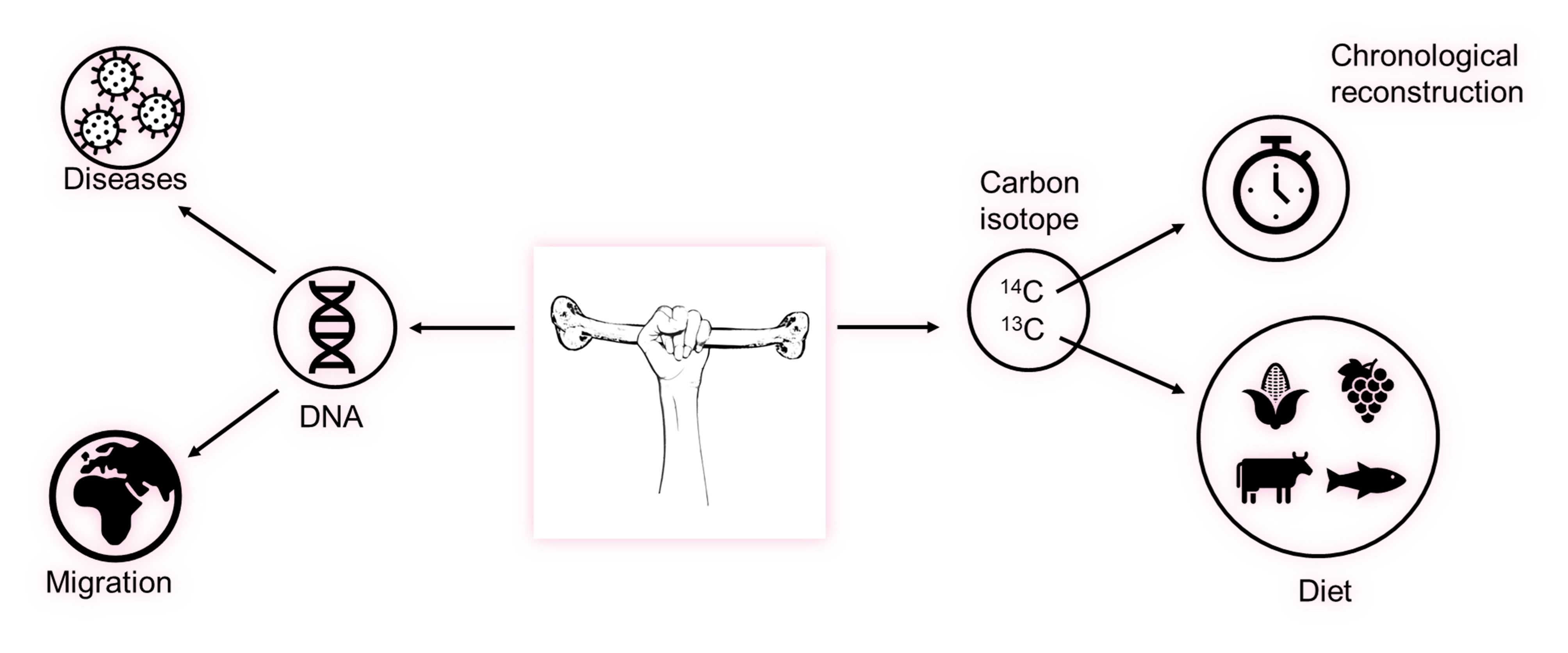
Archaeological bones are a sort of living memory from which we can glean a lot of information about ancient populations' lives: what they ate, their reproductive habits, the diseases that ravaged their bodies, and the migrations they undertook (Fig. 1). Timing the occurrences and changes of these behaviours are pivotal to understand human biological and cultural trajectories from an evolutionary perspective. A fundamental underpinning of these insights is chronology, with the radiocarbon (14C) 'clock' representing the backbone of chronological reconstructions for up to 55,000 years ago. However, sampling for 14C dating is a destructive process, and human fossils and bone artifacts are increasingly rare and more precious over time.

Figure 1.
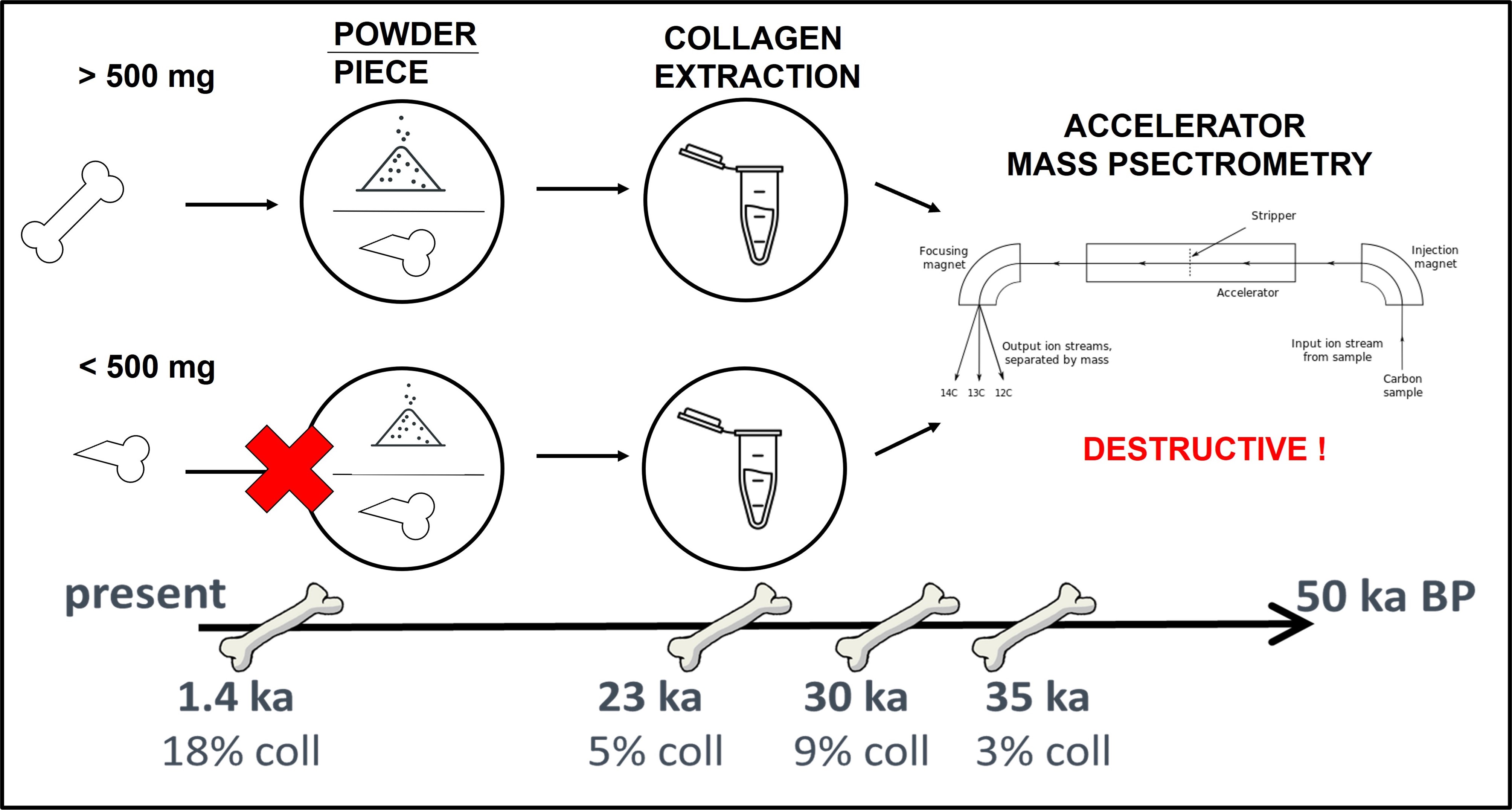
For proceeding with the 14C dating, a suitable amount of protein is requested, whose extraction includes, necessarily, a sampling of the bone itself. In light of these considerations, it is evident how this destructive step in dating protocols needs to be as less invasive as possible so that only bone fragments and areas with enough collagen are selected and submitted to the subsequent analysis (Fig. 2).
it is worth noting that preserving human and animal archaeological finds, with minimal invasive procedures, is not only a possibility but a duty of the scientific community. Nowadays, scientists involved in this research field must ensure that finds reach future generations of scientists, who will be able to apply more advanced strategies and tools for new discoveries about human evolution.

Figure 2.
Within this scenario, the advantage of spectroscopic images can be of high impact, in particular regarding the non-destructiveness and the possibility to map, from a spatial extent, the area of the bone in which the proteins are more present and to quantify if the collagen percentage is enough for 14C analysis.
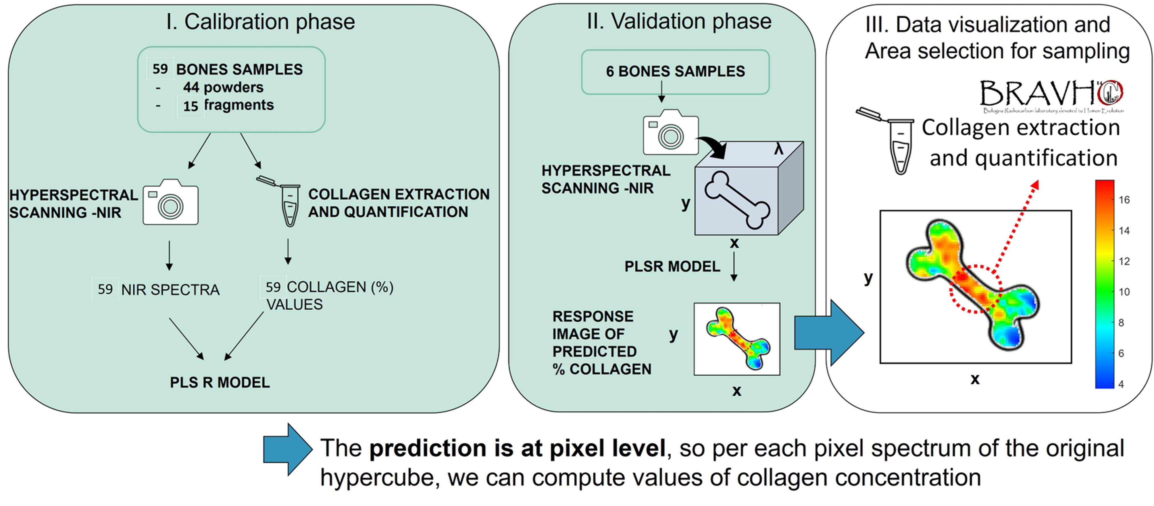
To this aim, a dedicated analytical protocol that combines hyperspectral cameras (working in the near-infrared region of the electromagnetic spectrum, referred to as HSI) with a tailored multivariate image analysis was studied to make the invisible visible, creating chemical images of the quantification and distribution of collagen in ancient bones (Fig. 3).

Figure 3.
The combination of different disciplines from analytical chemistry, chemometrics, archaeology, and radiocarbon dating was essential in creating an Italian research group dedicated to image analysis at the service of cultural heritage. This project was coordinated by the University of Bologna and the team from the University of Genova.
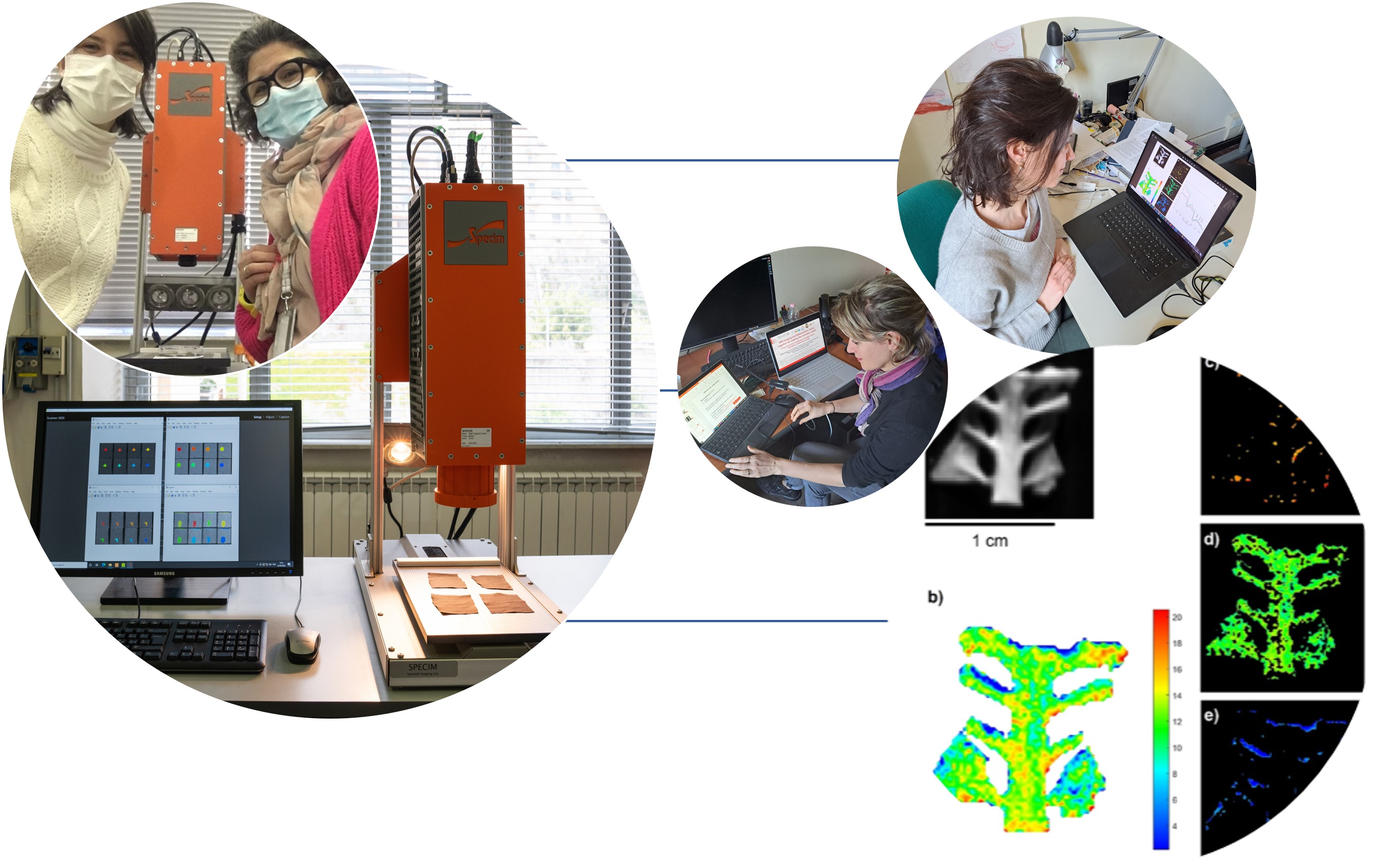
The interpretation of the results was always made in close cooperation between the two groups, merging the specific knowledges for drawing conclusions. This approach to scientific research is, in our opinion, essential for obtaining reliable methods and robust results and for contributing significantly to the growth of knowledge (Fig. 4).

Figure 4.
In more detail, the analytical procedure presented led to the construction of the quantitative model able to predict the percentage of collagen ascribable to every pixel of the image. To this aim, a set of samples, including both bone powders (44 samples) and fragments (15 samples), were analysed by means of HSI and subsequently submitted to collagen extraction, according to the most modern strategy developed in the BRAVHO Lab. These samples were chosen with the aim of covering a wide range of collagen percentages, from 0 to 20%, and with a focus on low collagen concentration, typical for prehistorical bones.
The mathematical calculation was performed modelling the correlations between the relative collagen amount and the NIR absorption spectra by means of a linear regression method (PLS – partial least squares), and subsequently, it was tested on 6 independent fragments. The collagen percentage of these 6 bones was unknown and it was determined only after image acquisition to confirm the model prediction ability. This rigid analytical protocol allows to obtain an error in collagen prediction equal to 2.2% (expressed ad RMSEP – root mean square error in prediction).
Ones the prediction of the amount of collagen present in the bone is performed, it is possible to build a chemical map of the analyte of interest (here the collagen) in which a false colour bar is used for representing areas with low (bluish colour) or high (reddish colour) percentage of collagen. Colours are also supported by the numerical prediction of the percentage itself so that the archaeologist can easily understand the real potential of an unknown samples for the collagen extraction and the consecutive 14C dating (Fig. 4).
Summarizing, this innovative and incisive combination of NIR-HSI spectroscopy prescreening and the radiocarbon method provides, for the first time, detailed information about the quantity of collagen on archaeological bones, as a crucial step forward for selecting the sampling point for 14C dating. This allows not only selecting the best specimens but also choosing the sampling point in the selected ones based on the amount of collagen predicted. This method helps to drastically reduce the number of samples destroyed for 14C analysis and within the bone, avoiding the selection of areas that may present a quantity of collagen not sufficient for the dating. With this work, we have shown that by using strategic coordination and effective research between spectroscopic methods and radiometric techniques to study prehistoric bones, we can provide a valuable contribution to the preservation, enhancement and protection of our cultural heritage.
Follow the Topic
-
Communications Chemistry

An open access journal from Nature Portfolio publishing high-quality research, reviews and commentary in all areas of the chemical sciences.
Related Collections
With Collections, you can get published faster and increase your visibility.
f-block chemistry
Publishing Model: Open Access
Deadline: Feb 28, 2026
Experimental and computational methodology in structural biology
Publishing Model: Open Access
Deadline: Apr 30, 2026


Please sign in or register for FREE
If you are a registered user on Research Communities by Springer Nature, please sign in