Production and use of antigen tetramers to study antigen-specific B cells
Published in Protocols & Methods, Immunology, and Anatomy & Physiology
We started developing this method with the goal of identifying rare antigen-specific naive B cells from non-transgenic mouse and human B cells when I started my postdoctoral research in Dr. Marc Jenkins’ Lab within the Center for Immunology at the University of Minnesota in January 2009. To my knowledge, at that time fewer than 100 studies had assessed antigen-specific B cells using flow cytometry since the first published use in 1972 (many of those reviewed here1). Many of those elegant studies focused upon transgenic B cells or a select number of model antigens. Additionally, previous studies often specifically excluded naïve B cells due to technical issues. In contrast, we wanted to analyze naïve and antigen-experienced B cells specific for any antigen of interest. When I joined the Jenkins Lab, a talented scientist Dr. Kathy Pape had developed a protocol to enrich and analyze rare B cells specific for fluorescent model antigens2 built upon other enrichment approaches3-5 that I aimed to modify. Initially I conjugated a fluorochrome directly to the model antigen ovalbumin following direct labeling approaches utilized by others in the field6. Unfortunately, while I could detect ovalbumin-specific B cells using flow cytometry with this tool, it was clear that a lot of the naïve B cells able to respond to ovalbumin immunization were missed. The failure of our first approach was not entirely unexpected given the rising use of multimerized probes to stabilize low affinity binding, but it gave us a sense of just how much of the relevant B cell repertoire was missed with a monomeric probe. We next began using antigen tetramers to detect antigen-specific B cells, which to my knowledge had been used by only a few studies when the project began1,7-9, and a few more10-13 before we published in 201214.
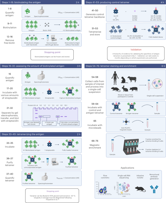
The tetramer approach had a few technical hurdles. The first hurdle was biotinylation. Typically, protocols are set up to attach multiple biotins to a protein, but we were concerned that this approach would result in aggregation of our detection tool if multiple streptavidin molecules bound to biotins on the same antigen molecule. To avoid this, we went low tech and performed our conjugation reactions at suboptimal concentrations such that there were fewer biotin molecules than antigen molecules, thereby decreasing the probability of multiple biotins on an antigen molecule. To assess the level of biotinylation we adapted a protocol Dr. Jim Moon and others in the Jenkins lab used to assess biotinylation of the peptide:MHC molecules used to create tetramers for assessing antigen-specific T cells15,16. Essentially that protocol just made little batches of tetramer at various antigen to streptavidin ratios and assessed for the point at which biotinylated molecules were confirmed to be in excess.
A second hurdle was that we knew there would be B cells specific for the fluorochrome in the tetramer17-19 and suspected that there would be B cells specific for streptavidin. We didn’t publish the data until this 2024 Nature Protocols paper, but one can demonstrate this nicely as an expansion of tetramer-binding B cells after immunization with fluorochrome or streptavidin in adjuvant. We’ve also shown that antibodies specific for tags included in recombinant proteins can bind to tetramers20. In our original work we did show that the frequency of B cells binding the streptavidin-fluorochrome conjugate was higher than the frequency of B cells specific for our antigen of interest14, meaning that just looking at all B cells binding antigen tetramers would be mostly cells that were irrelevant to our study.
Previously, an elegant approach to exclude fluorochrome-specific B cells was developed which co-labeled with the same antigen bound to two different fluorochromes21. Unfortunately, using this approach streptavidin-specific and tag-specific B cells would contaminate the antigen-specific population since they would also be co-labeled. Instead of adding a third tetramer to exclude B cells specific for streptavidin/tag, we developed a very simple control (aka “decoy”) tetramer which would allow us to gate out any cells of unwanted specificity binding to our antigen tetramer. We simply added Alexa Fluor 647 (AF647) to streptavidin-PE, creating streptavidin-PE-AF647 which created FRET and could be robustly detected in the channels most often used for PE-Cy5 or PerCP-Cy5.5.
Questions I’m often asked about this tool:
Why AF647 and not other fluors?
A few reasons:
- I found emission and excitation info for PE-AF647, but not every other possible combination.
- I tried PE-AF750 but the fluorescence was fairly dim. Admittedly, I didn’t try to optimize this because I didn’t want to give up the PE-Cy7 channel for a control reagent.
- PE-Cy5 was an underutilized channel on LSRII machines in those days with most people avoiding it completely due to spill and spreading issues with other channels.
- DyLight 650 works just as well, as does DyLight 594.
Why make your own conjugates instead of commercially available ones?
When I was exploring ovalbumin directly conjugated to PE I realized that PE (R-Phycoerythrin) from different companies was different and these changes altered some of the epitopes available for B cells to bind. Essentially, if I co-labeled with PE and PE-AF647 I made using PE from the same company I’d co-label all of the cells that bound PE. If I did the same experiment using mismatched PE from different companies, I would detect a population of B cells that bound PE but not PE-AF647. After this experience I realized that company-to-company differences could sink my experiments, so I needed to be extremely careful with my tools if I didn’t want to analyze artifacts.
Your follow-up question is probably: Why not just buy streptavidin-PE and streptavidin-PE-Cy5 from the same company?
The first reason was that I was afraid that I didn’t know the chemistry used by companies and would rather trust empirical evidence. The second reason some unpublished evidence from the Jenkins Lab and the NIH tetramer core at Emory that indicated that streptavidin-PE from Prozyme (later bought out by Agilent) better separation to noise compared to streptavidin-PE offered by other companies at the time. (Note: The NIH tetramer core by MHC tetramer pioneer Dr. John Altman22 still uses this streptavidin-PE, unclear if anyone has done additional head-to-head tests).
Along the way this became a really great collaboration with Dr. Ryan Martinez from Dr. Dan Mueller’s lab that was next door to the Jenkins Lab. Ryan was a technician at the time we handed the project back and forth after I developed the “decoy” approach, and I ended up finishing it with help from other members of the Mueller Lab when Ryan went on to get his MD/PhD from Emory University. Most prominently, another talented technician from the Mueller Lab Dr. Phil Titcombe, who later obtained his PhD from University of Minnesota. Ryan really refined the strategy and brought a whole new dynamic to the paper adding most of the GPI experiments. We ended up using these approaches to assess B cells specific self-antigens and sensitively quantitated deletion and anergy in these models which classically had been done using elegant BCR transgenic models. Many of our findings have been reproduced and extended by other groups, which is always satisfying to see. I’ve gotten great joy out of handing these protocols off to other labs led by Dr. Marion Pepper, Dr. James McLachlan, Dr. Camila Coelho, Dr. Jim Boonyaratanakornkit, Dr. Marco Pravetoni, and Dr. Josh Koenig, just to name a few who continue to use the approach. Josh posted about his experience here. He deserves a lot of credit for leading the writeup of this protocol, and pushing me to write this post.
I also want to mention that while this project was a success, it grew out of a failed attempt to study human Hepatitis B-specific B and T cells before and after vaccination, which was my initial project as a postdoc. This project was the focus of my 2012 Irvington Fellowship from the Cancer Research Institute, but a quick scan of my publication record will reveal that I’ve never published anything on Hepatitis B, or anything using human samples from my time as a postdoctoral researcher. Fortunately, I had the support and freedom to pivot that failure towards something more fruitful that ultimately allowed me to start my lab in 2014 at the Fred Hutchinson Cancer Center and these protocols are critical for most of the projects my lab will work on for years to come at the Beirne B. Carter Center for Immunology Research at the University of Virginia.
- Moody MA, Haynes BF. Antigen-specific B cell detection reagents: use and quality control. Cytometry A. 2008;73(11):1086-92. Epub 2008/07/10. doi: 10.1002/cyto.a.20599. PMID: 18613115.
- Pape KA, Taylor JJ, Maul RW, Gearhart PJ, Jenkins MK. Different B cell populations mediate early and late memory during an endogenous immune response. Science. 2011;331(6021):1203-7. Epub 2011/02/12. doi: science.1201730 [pii]
10.1126/science.1201730. PMID: 21310965.
- Irsch J, Hunzelmann N, Tesch H, Merk H, Maggi E, Ruffilli A, Radbruch A. Isolation and characterization of allergen-binding cells from normal and allergic donors. Immunotechnology. 1995;1(2):115-25. doi: 10.1016/1380-2933(95)00012-7. PMID: 9373340.
- Leyendeckers H, Odendahl M, Lohndorf A, Irsch J, Spangfort M, Miltenyi S, Hunzelmann N, Assenmacher M, Radbruch A, Schmitz J. Correlation analysis between frequencies of circulating antigen-specific IgG-bearing memory B cells and serum titers of antigen-specific IgG. Eur J Immunol. 1999;29(4):1406-17. doi: 10.1002/(SICI)1521-4141(199904)29:04<1406::AID-IMMU1406>3.0.CO;2-P. PMID: 10229109.
- Martin F, Oliver AM, Kearney JF. Marginal zone and B1 B cells unite in the early response against T-independent blood-borne particulate antigens. Immunity. 2001;14(5):617-29. doi: 10.1016/s1074-7613(01)00129-7. PMID: 11371363.
- Hoven MY, De Leij L, Keij JF, The TH. Detection and isolation of antigen-specific B cells by the fluorescence activated cell sorter (FACS). J Immunol Methods. 1989;117(2):275-84. doi: 10.1016/0022-1759(89)90150-6. PMID: 2784157.
- Newman J, Rice JS, Wang C, Harris SL, Diamond B. Identification of an antigen-specific B cell population. J Immunol Methods. 2003;272(1-2):177-87. doi: 10.1016/s0022-1759(02)00499-4. PMID: 12505722.
- Mulder A, Eijsink C, Kardol MJ, Franke-van Dijk ME, van der Burg SH, Kester M, Doxiadis, II, Claas FH. Identification, isolation, and culture of HLA-A2-specific B lymphocytes using MHC class I tetramers. J Immunol. 2003;171(12):6599-603. doi: 10.4049/jimmunol.171.12.6599. PMID: 14662862.
- Zachary AA, Kopchaliiska D, Montgomery RA, Leffell MS. HLA-specific B cells: I. A method for their detection, quantification, and isolation using HLA tetramers. Transplantation. 2007;83(7):982-8. doi: 10.1097/01.tp.0000259017.32857.99. PMID: 17460571.
- Han M, Rogers JA, Lavingia B, Stastny P. Peripheral blood B cells producing donor-specific HLA antibodies in vitro. Hum Immunol. 2009;70(1):29-34. Epub 20081119. doi: 10.1016/j.humimm.2008.10.013. PMID: 19026703.
- Verkoczy L, Moody MA, Holl TM, Bouton-Verville H, Scearce RM, Hutchinson J, Alam SM, Kelsoe G, Haynes BF. Functional, non-clonal IgMa-restricted B cell receptor interactions with the HIV-1 envelope gp41 membrane proximal external region. PLoS One. 2009;4(10):e7215. Epub 20091006. doi: 10.1371/journal.pone.0007215. PMID: 19806186; PMCID: PMC2751816.
- Franz B, May KF, Jr., Dranoff G, Wucherpfennig K. Ex vivo characterization and isolation of rare memory B cells with antigen tetramers. Blood. 2011;118(2):348-57. Epub 20110506. doi: 10.1182/blood-2011-03-341917. PMID: 21551230; PMCID: PMC3138687.
- Morris L, Chen X, Alam M, Tomaras G, Zhang R, Marshall DJ, Chen B, Parks R, Foulger A, Jaeger F, Donathan M, Bilska M, Gray ES, Abdool Karim SS, Kepler TB, Whitesides J, Montefiori D, Moody MA, Liao HX, Haynes BF. Isolation of a human anti-HIV gp41 membrane proximal region neutralizing antibody by antigen-specific single B cell sorting. PLoS One. 2011;6(9):e23532. Epub 20110930. doi: 10.1371/journal.pone.0023532. PMID: 21980336; PMCID: PMC3184076.
- Taylor JJ, Martinez RJ, Titcombe PJ, Barsness LO, Thomas SR, Zhang N, Katzman SD, Jenkins MK, Mueller DL. Deletion and anergy of polyclonal B cells specific for ubiquitous membrane-bound self-antigen. The Journal of experimental medicine. 2012;209(11):2065-77. doi: 10.1084/jem.20112272. PMID: 23071255; PMCID: 3478923.
- Moon JJ, Chu HH, Pepper M, McSorley SJ, Jameson SC, Kedl RM, Jenkins MK. Naive CD4(+) T cell frequency varies for different epitopes and predicts repertoire diversity and response magnitude. Immunity. 2007;27(2):203-13. Epub 20070816. doi: 10.1016/j.immuni.2007.07.007. PMID: 17707129; PMCID: PMC2200089.
- Moon JJ, Chu HH, Hataye J, Pagan AJ, Pepper M, McLachlan JB, Zell T, Jenkins MK. Tracking epitope-specific T cells. Nat Protoc. 2009;4(4):565-81. doi: 10.1038/nprot.2009.9. PMID: 19373228; PMCID: PMC3517879.
- Maruyama M, Lam KP, Rajewsky K. Memory B-cell persistence is independent of persisting immunizing antigen. Nature. 2000;407(6804):636-42. doi: 10.1038/35036600. PMID: 11034213.
- Hayakawa K, Ishii R, Yamasaki K, Kishimoto T, Hardy RR. Isolation of high-affinity memory B cells: phycoerythrin as a probe for antigen-binding cells. Proceedings of the National Academy of Sciences of the United States of America. 1987;84(5):1379-83. doi: 10.1073/pnas.84.5.1379. PMID: 3493491; PMCID: PMC304433.
- Bell J, Gray D. Antigen-capturing cells can masquerade as memory B cells. The Journal of experimental medicine. 2003;197(10):1233-44. doi: 10.1084/jem.20020270. PMID: 12756262; PMCID: PMC2193792.
- Fitzpatrick KS, Degefu HN, Poljakov K, Bibby MG, Remington AJ, Searles TG, Gray MD, Boonyaratanakornkit J, Rosato PC, Taylor JJ. Validation of Ligand Tetramers for the Detection of Antigen-Specific Lymphocytes. J Immunol. 2023;210(8):1156-65. doi: 10.4049/jimmunol.2200934. PMID: 36883850; PMCID: PMC10073333.
- Townsend SE, Goodnow CC, Cornall RJ. Single epitope multiple staining to detect ultralow frequency B cells. J Immunol Methods. 2001;249(1-2):137-46. doi: 10.1016/s0022-1759(00)00352-5. PMID: 11226471.
- Altman JD, Moss PA, Goulder PJ, Barouch DH, McHeyzer-Williams MG, Bell JI, McMichael AJ, Davis MM. Phenotypic analysis of antigen-specific T lymphocytes. Science. 1996;274(5284):94-6. doi: 10.1126/science.274.5284.94. PMID: 8810254.
Immunologist specializing in B cell analysis and engineering
@justintaylorlab
@justintaylorlab.bsky.social
Follow the Topic
-
Nature Protocols
This journal publishes secondary research articles and covers new techniques and technologies, as well as established methods, used in all fields of the biological, chemical and clinical sciences.

Ask the Editor - Immunology, Pathogenesis, Inflammation and Innate Immunity
Got a question for the editor about the complement system in health and disease? Ask it here!
Continue reading announcement


Please sign in or register for FREE
If you are a registered user on Research Communities by Springer Nature, please sign in